
Skip to main content Skip to main navigation menu Skip to site footer
logo
  • Login
  • Register
E-ISSN: 2822-6453
  • Home
  • Current Issue
  • Archives
  • For Authors
    • Author Guidelines
    • Online Submission
    • Reference Style
    • Call for Papers
    • Language Editing
  • For Reviewers
    • Reviewer Guidelines
    • Become a Reviewer
    • Reviewer Resources
  • Ethics and Policies
    • Artificial Intelligence (AI) Policy
    • Authorship and Contributorship
    • Conflicts of Interest
    • Copyright and Permission
    • Corrections and Retractions
    • Crossmark Policy
    • Editorial Policy
    • Human and Animal Rights
    • Informed Consent Policy
    • Journal Management
    • Open Access Statement
    • Publication Ethics Policy
    • Research Ethics Policy
  • Journal Info
    • About the Journal
    • Abstracting & Indexing
    • Editorial Board
    • Peer Review Process
    • Publication Fee
    • Society Information
    • Contact
  1. Home /
  2. Search

Search

Advanced filters
Published After
Published Before

Search Results

##search.searchResults.foundPlural##
  • Full-arch dental impressions: Conventional, digital, and photogrammetric techniques

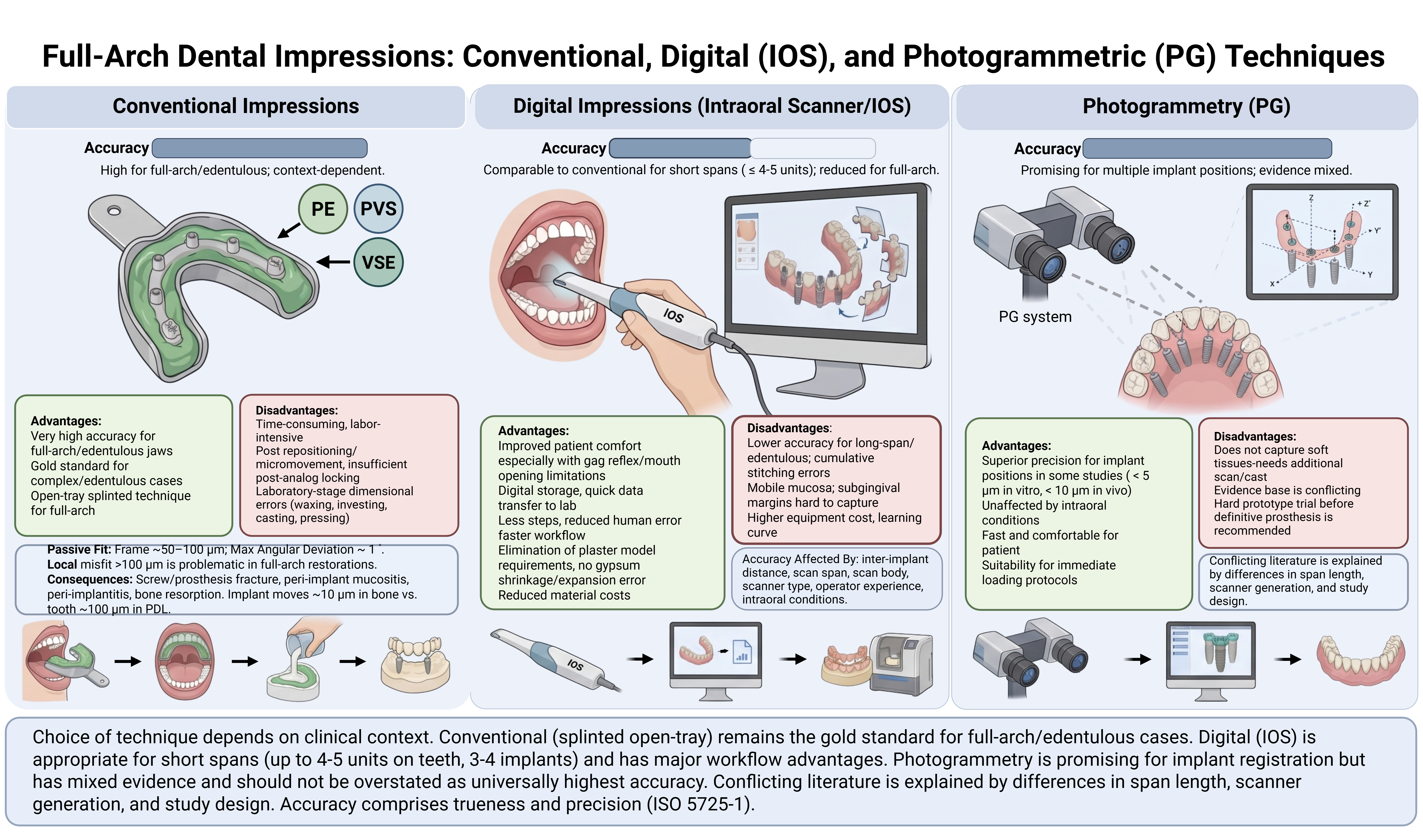
    Advances in digital dentistry now permit the fabrication of full-arch prosthetic restorations through a fully digital workflow, extending from impression acquisition to the production of the definitive prosthesis. Intraoral scanning has become an acceptable alternative to conventional impression techniques, providing valuable information for diagnosis, restorative dentistry, orthodontic analysis, and implant-supported prosthodontics. Despite the many advantages offered by digital methods, conventional impression techniques using polyether, polyvinyl siloxane, and vinyl siloxanether materials remain widely used by clinicians because of their excellent dimensional stability and well-documented precision. This review compares and critically discusses conventional and digital impression techniques for acquiring full-arch dental registrations, with particular attention to their accuracy, advantages, limitations, and clinical indications. The accuracy of digital impressions is strongly influenced by inter-implant distance, scan span length, scan body geometry and material, scanner type, operator experience, and intraoral conditions such as mobile mucosa, saliva, and limited anatomical landmarks. For single-unit and short-span restorations of up to four or five units, intraoral scanning achieves accuracy comparable to that of conventional techniques. In long-span and full-arch restorations, however, accuracy tends to decline owing to cumulative image-stitching errors and the scarcity of reference points, a problem that is especially pronounced in completely edentulous arches. Photogrammetry has emerged as a promising alternative for the reliable registration of multiple implant positions, although it inadequately captures peri-implant soft tissues. Overall, technique selection depends primarily on the number of teeth or implants and the distance between them; conventional methods remain preferable for completely edentulous patients, whereas digital workflows are suitable for restorations spanning up to three or four implants. Despite persisting limitations, digital techniques are strong candidates to eventually outperform conventional approaches in dentistry.
    Full-arch impression, conventional impression, digital impression, intraoral scanner, photogrammetry, implant-supported prosthesis, impression accuracy
    Ebru Sümer Ekin
    27.08.2026
    e260826
    Abstract Views: 430 Download: 142 DOI 10.5577/jomdi.e260826
  • Can digital impression methods eliminate the current problems of conventional impression techniques?

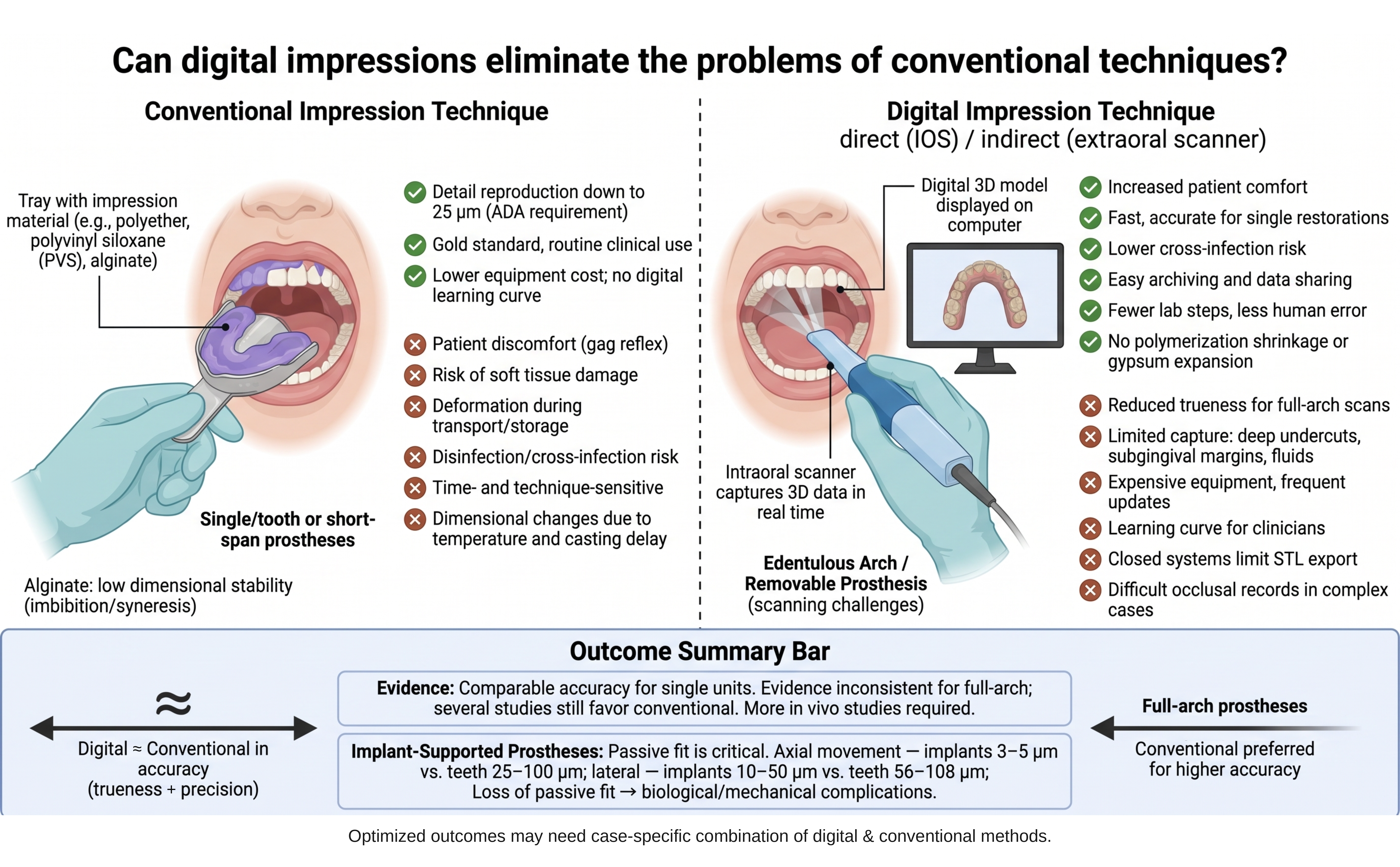
    Conventional impression materials remain a cornerstone of routine dental practice, yet they carry well-documented drawbacks, including a pronounced gag reflex, limited patient comfort, potential soft-tissue trauma, dimensional changes with temperature and elapsed time, the need for disinfection, cross-infection risk, and susceptibility to distortion during transport, storage, and laboratory handling. Digital impression systems have been introduced to overcome these limitations, and their use in dentistry—particularly in prosthodontics—continues to expand. This review compares conventional and digital impression techniques and examines whether digital workflows can resolve the shortcomings inherent to conventional methods. Conventional elastomers such as polyether and addition-type silicones offer excellent dimensional stability and detail reproduction and are currently regarded as the gold standard. Digital impressions, obtained with intraoral or extraoral scanners, provide clear advantages in patient comfort, procedure time, reduced cross-infection risk, elimination of polymerization shrinkage and gypsum expansion, and convenient digital archiving and transfer. However, their accuracy is influenced by scanning area, scanner sensitivity, operator experience, moisture, patient movement, and inter-implant distance. The reviewed literature indicates that intraoral scanners achieve clinically acceptable accuracy for single units and single-quadrant restorations, whereas evidence for full-arch and multiple implant-supported prostheses remains inconsistent, with several studies still favoring conventional impressions. Accuracy—comprising trueness and precision—is especially critical for passive fit in implant-supported restorations. Most comparative data derive from in vitro models that do not reproduce intraoral conditions such as saliva, blood, and patient movement, underscoring the need for further in vivo research. In conclusion, although the benefits of digital impressions are evident, their long-term clinical efficacy and safety require additional investigation, and technique selection should weigh accuracy, time efficiency, learning curve, and cost. A case-specific combination of digital and conventional approaches may offer added benefit.
    Digital impression, intraoral scanner, conventional impression, trueness and precision, prosthodontics
    Yakup Kantaci
    27.08.2026
    e260815
    Abstract Views: 387 Download: 170 DOI 10.5577/jomdi.e260815
  • An overview of intraoral scanners and computer-aided design software programs used in scan design stages in digital dentistry

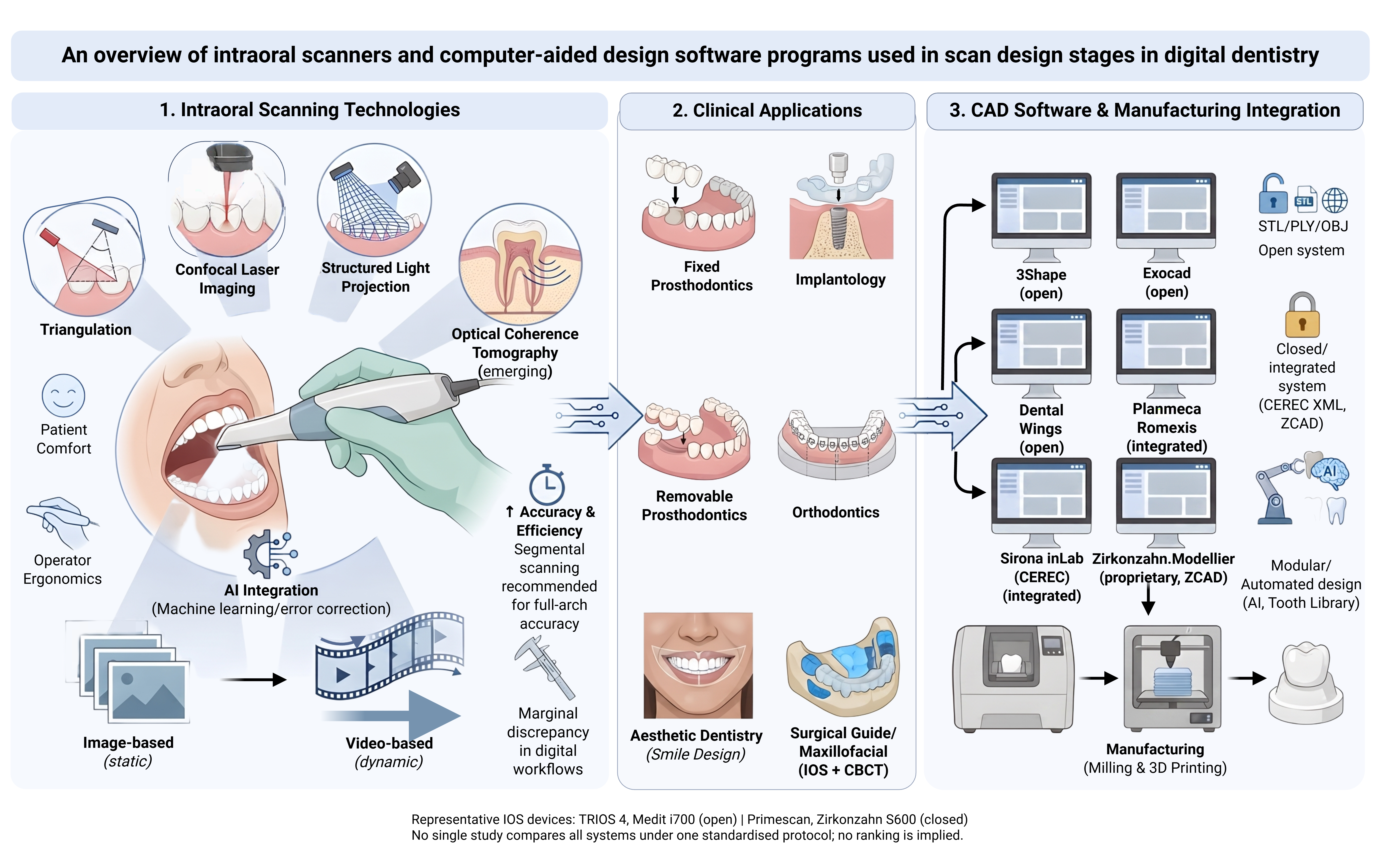
    In prosthetic dental treatments, obtaining the negative of the prepared area in the most accurate way directly affects the success of the restoration to be made. Therefore, it is necessary to give importance to the impression material and the impression technique used. Impression materials have undergone changes over the years in order to record the most accurate and sensitive detail. Studies have tried to improve many physical and chemical properties of the impression material. Despite this, it should not be forgotten that there are numerous external factors such as the skill of the clinician, environmental conditions, and storage conditions for the impression to be obtained. Developments in computer-aided design and design (CAD/CAM) technology have significantly increased the use of these systems in dentistry in recent years. In dentistry, many companies in the market aim to minimize the problems that may occur in the laboratory and clinical environment by adding intraoral or extraoral scanners to CAD/CAM systems. The first step of dental CAD-CAM systems is digital scanning with an intraoral or laboratory scanner. This step is followed by the construction phase of restorations with digital design and production devices on the computer. Therefore, the success of these restorations depends largely on the accuracy and precision of the scanning devices. This review will discuss CAD/CAM systems and intraoral scanners and software programs.
    Digital dentistry, intraoral scanners, CAD/CAM systems, digital impression, scanning accuracy, digital workflow
    Muhammed Burak Süzen, Hayriye Yasemin Yay Kuşçu
    27.08.2026
    e260828
    Abstract Views: 538 Download: 184 DOI 10.5577/jomdi.e260828
  • How should I scan for measurement accuracy in full-arch scans?

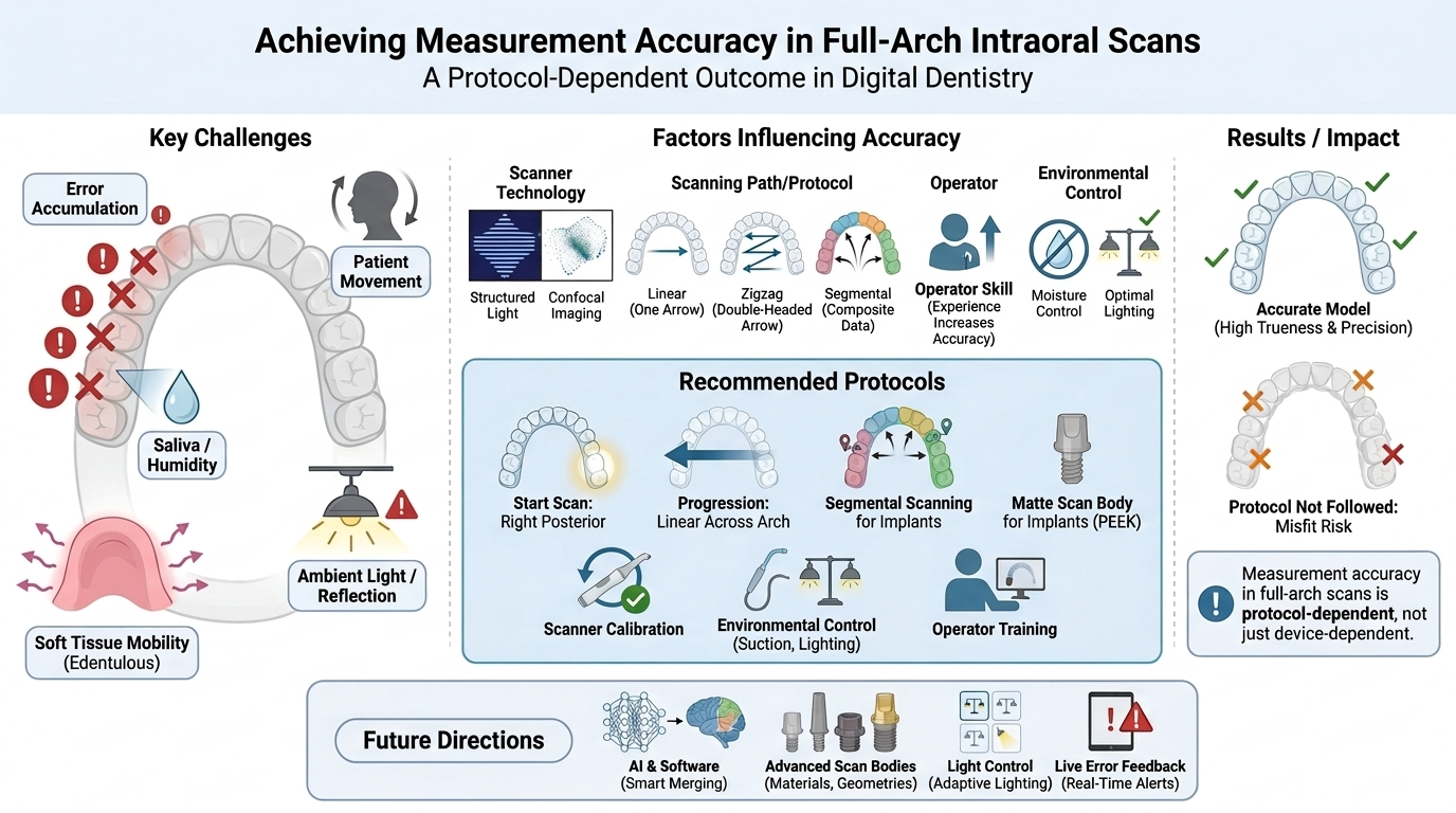
    The integration of intraoral scanners (IOSs) into routine clinical practice has transformed full-arch digital impression-making, offering improved patient comfort and the potential for high measurement accuracy. However, ensuring reliable trueness and precision across large arch spans remains challenging because of the cumulative errors that arise when extended areas are scanned, which can compromise the fit of prosthetic restorations, generate stress within implant-supported frameworks, and introduce clinically relevant errors in orthodontic planning. This review synthesizes the current evidence on the technical, biological, and operator-related factors that influence full-arch scanning accuracy and outlines clinically applicable strategies to minimize error. The principal challenges discussed include local error accumulation, patient movement, saliva and moisture, soft-tissue mobility in edentulous arches, and operator fatigue, all of which can lead to volumetric deformation of the digital model. Key determinants of accuracy are examined, including scanner technology, scanning path and starting-point selection, operator experience, oral humidity, ambient light, and implant scan-body characteristics. Recommended scanning patterns—linear, zigzag, and segmental approaches—are compared and discussed, together with region-specific strategies for the anterior and posterior segments. The evidence indicates that a disciplined protocol markedly improves trueness and precision: linear progression from the posterior toward the anterior region, segmental scanning in implant-supported cases, regular scanner calibration, a constant scanning speed and distance, effective moisture and ambient-light control, the use of matte scan bodies such as PEEK, and continuous operator training. Future research should prioritize intelligent auto-alignment algorithms, standardized implant scan-body geometries and materials, multicenter evaluation of environmental conditions, and artificial-intelligence-based real-time error detection to further enhance the reliability of digital impressions in clinical practice.
    Full-arch scanning, intraoral scanner, trueness and precision, segmental scanning, digital impression
    Savaş Sağmak, Muhammed Burak Süzen
    27.08.2026
    e260829
    Abstract Views: 342 Download: 131 DOI 10.5577/jomdi.e260829
  • Comparison of scanning accuracy of different digital intraoral scanners

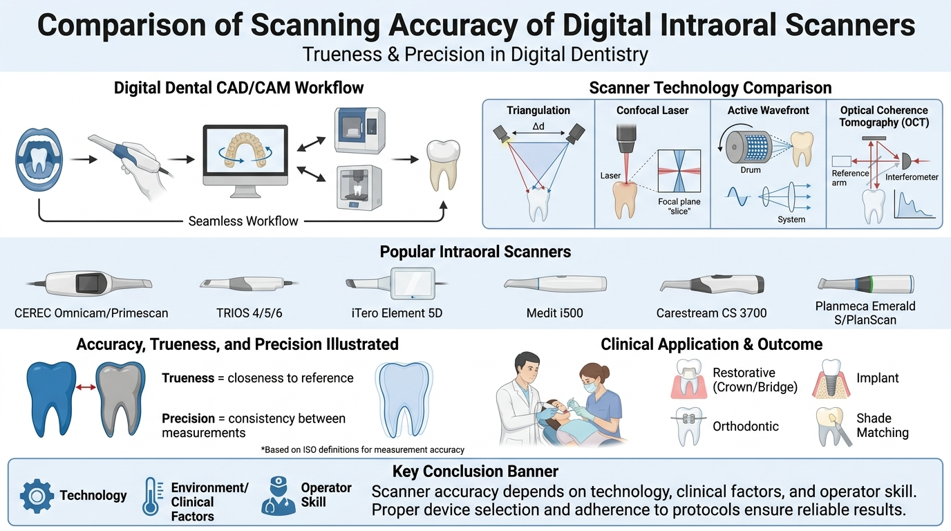
    Digital intraoral scanners (IOSs) have gained significant importance in modern dentistry by offering accurate, efficient, and patient-friendly alternatives to conventional impression techniques. This review aims to evaluate and compare the image quality and accuracy provided by various intraoral scanners, focusing on the current literature. Factors influencing image quality, such as scanning technology (confocal microscopy, triangulation, active wavefront sampling), operator experience, and scanning strategy, are also discussed. The evaluation includes commonly used IOS devices, including iTero, Trios, Medit i700, and CEREC Primescan, analyzing their performance in clinical and laboratory conditions. The literature suggests that while each scanner has strengths and limitations, all of them provide clinically acceptable results for prosthodontic and orthodontic applications. Furthermore, advancements in software and hardware are continuously enhancing image resolution, data acquisition speed, and ease of use. However, despite the progress, discrepancies in trueness and precision still exist depending on the scanning environment, scan area size, and material reflectivity. This review concludes that selecting an appropriate IOS should be based on clinical needs, operator preferences, and the specific task at hand. Continuous technological development and standardized evaluation methods are essential to further improve IOS reliability and integration into routine clinical practice.
    Accuracy, intraoral scanners, digital scanning, three-dimensional imaging, digital dentistry
    Elif Atasever, Server Mutluay Ünal
    27.08.2026
    e260823
    Abstract Views: 379 Download: 130 DOI 10.5577/jomdi.e260823
1 - 5 of 5 items

Journal Issue

Vol. 7 No. S1 (2026) Continuous Publication
View Current Issue

Published Volumes

2026
  • Vol. 7 No. S1 (2026): Current Developments in Prosthetic Dentistry
  • Vol. 7 (2026): Journal of Medical and Dental Investigations
2025
  • Vol. 6 No. S1 (2025): Current Developments in Pediatric Dentistry
  • Vol. 6 No. Advanced Online (2025): Journal of Medical and Dental Investigations
  • Vol. 6 (2025): Journal of Medical and Dental Investigations
2024
  • Vol. 5 (2024): Journal of Medical and Dental Investigations
2023
  • Vol. 4 (2023): Journal of Medical and Dental Investigations
2022
  • Vol. 3 (2022): Journal of Medical and Dental Investigations
Show more Show less
2021
  • Vol. 2 (2021): Journal of Medical and Dental Investigations

socialshare

Submit Your Paper
Become a Reviewer

sidebarmenu

Journal Menu

 Home

 Aims & scopes

 Editorial Board

 Author Guidelines

 Abstracting & Indexing

 Peer Review Process

 Ethics Statement

 Plagiarism Policy

 Digital Archiving Policy

 Corrections and Retractions

 Journal History

 Journal Statistics

 Society Information

 Language Editing

 Contact

idr


Citations
Updated daily Google Scholar
All Since 2021
Citations 44 44
h-index 4 4
i10-index 0 0
0 5 10 15 20 25
  • 2 2023
  • 7 2024
  • 21 2025
  • 14 2026
Citations per year
Year Citations
2023 2
2024 7
2025 21
2026 (to date) 14

2026 counts citations recorded so far this year.

ithenticate

Indexed in CiteLink


Signatory of DORA

Latest publications

  • Atom logo
  • RSS2 logo
  • RSS1 logo

Information

  • For Readers
  • For Authors
  • For Librarians

Browse

  • Categories
    • DENTISTRY
    • MEDICINE
    • Open Access
    • Original Article
    • Review
    • Case Report
    • Letter to Editor
Most Read Articles
  • Restorative options for primary teeth in pediatric dentistry
    3509 3509 views 2025
  • Use of platelet-rich fibrin (PRF) in the periodontology: A review
    3059 3059 views 2023
  • Endodontic treatment in primary teeth
    2296 2296 views 2025
  • Potential complications and preventive measures in pediatric dental treatments performed under general anesthesia and sedation
    2227 2227 views 2025
  • Does a sloped neck implant design facilitate All-on-4 surgeries? A three-dimensional finite element analysis
    2213 2213 views 2023
Most Cited Articles
  • Use of platelet-rich fibrin (PRF) in the periodontology: A review
    7 7 citations 2023
  • Evaluation of time exposure of different irrigation agents on radicular dentin erosion
    7 7 citations 2022
  • Comparison of second mesiobuccal canal prevalence of maxillary molar teeth by gender and age in a Turkish subpopulation: A cone-beam computed tomography study
    4 4 citations 2021
  • Does a sloped neck implant design facilitate All-on-4 surgeries? A three-dimensional finite element analysis
    4 4 citations 2023
  • Effect of different polishing methods applied to denture base materials on microbial adhesion and surface roughness
    4 4 citations 2023

visitors

Visitors 

Flag Counter

Web Analytics Made Easy - Statcounter

View My Stats

announcement

Announcement

logo
E-ISSN: 2822-6453

Download App

Contact

Publisher: Journal of Medical and Dental Investigations

Email: info@jomdi.org

Website: www.jomdi.org

Address: Publisher ZEUS PUBLISHING www.zeuspublishing.com

Social Media

CiteLinkEBSCOOpen AccessCrossrefGoogle ScholarCOPECreative Commons

Copyright © 2020-2026 | Journal of Medical and Dental Investigations. All rights reserved.
CC BY Open Access articles are distributed under the terms of the Creative Commons Attribution 4.0 International License,
which permits unrestricted use, distribution, and reproduction provided the original work is properly cited.

Privacy notice

This journal stores a session cookie that is required for the site to work, and keeps usage statistics that do not identify you.

More information