
 10.5577/jomdi.e260829
Vol. 7 No. S1 (2026) Continuous Publication
View Current Issue
Search
Search Results
##search.searchResults.foundPlural##
-
How should I scan for measurement accuracy in full-arch scans?
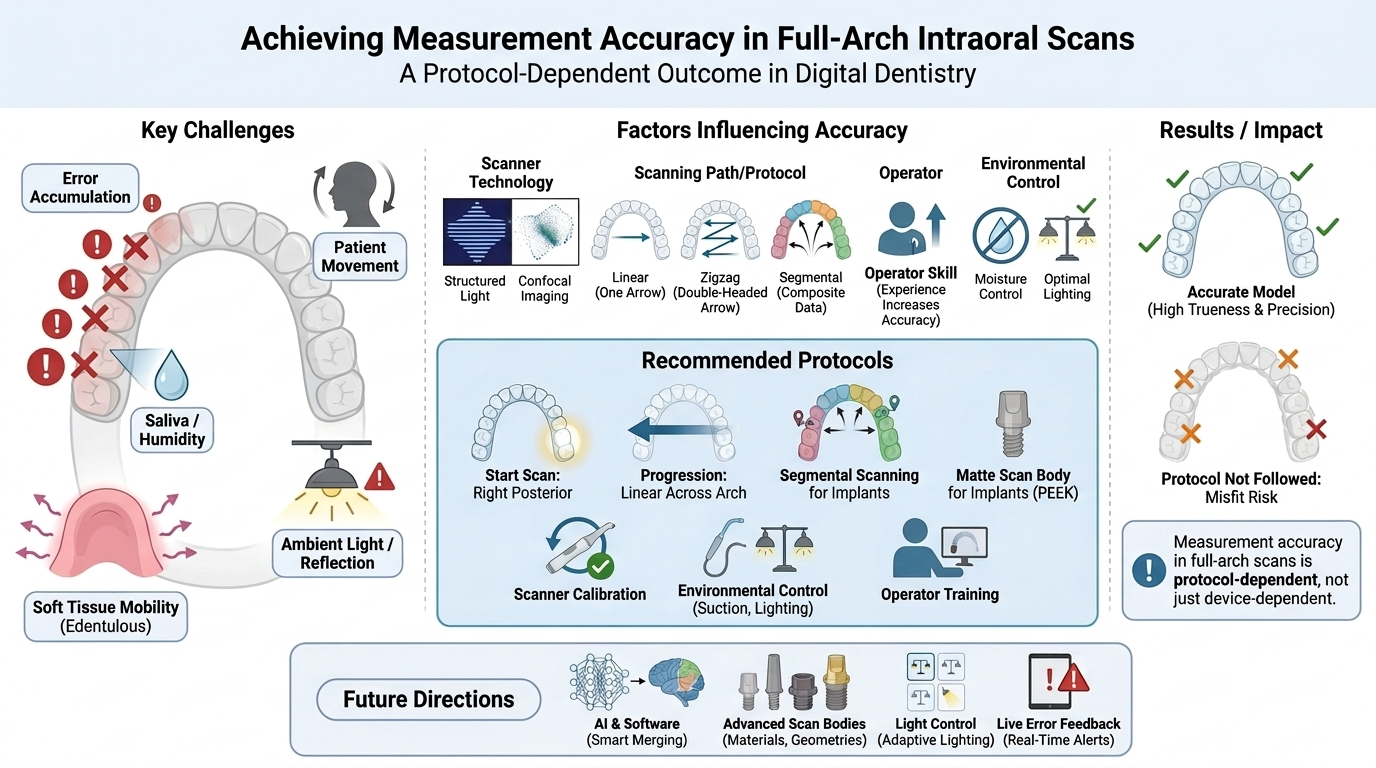
The integration of intraoral scanners (IOSs) into routine clinical practice has transformed full-arch digital impression-making, offering improved patient comfort and the potential for high measurement accuracy. However, ensuring reliable trueness and precision across large arch spans remains challenging because of the cumulative errors that arise when extended areas are scanned, which can compromise the fit of prosthetic restorations, generate stress within implant-supported frameworks, and introduce clinically relevant errors in orthodontic planning. This review synthesizes the current evidence on the technical, biological, and operator-related factors that influence full-arch scanning accuracy and outlines clinically applicable strategies to minimize error. The principal challenges discussed include local error accumulation, patient movement, saliva and moisture, soft-tissue mobility in edentulous arches, and operator fatigue, all of which can lead to volumetric deformation of the digital model. Key determinants of accuracy are examined, including scanner technology, scanning path and starting-point selection, operator experience, oral humidity, ambient light, and implant scan-body characteristics. Recommended scanning patterns—linear, zigzag, and segmental approaches—are compared and discussed, together with region-specific strategies for the anterior and posterior segments. The evidence indicates that a disciplined protocol markedly improves trueness and precision: linear progression from the posterior toward the anterior region, segmental scanning in implant-supported cases, regular scanner calibration, a constant scanning speed and distance, effective moisture and ambient-light control, the use of matte scan bodies such as PEEK, and continuous operator training. Future research should prioritize intelligent auto-alignment algorithms, standardized implant scan-body geometries and materials, multicenter evaluation of environmental conditions, and artificial-intelligence-based real-time error detection to further enhance the reliability of digital impressions in clinical practice.Full-arch scanning, intraoral scanner, trueness and precision, segmental scanning, digital impressionSavaş Sağmak, Muhammed Burak Süzen27.08.2026e260829 -
Comparison of scanning accuracy of different digital intraoral scanners
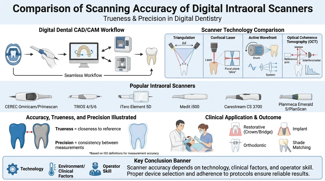
Digital intraoral scanners (IOSs) have gained significant importance in modern dentistry by offering accurate, efficient, and patient-friendly alternatives to conventional impression techniques. This review aims to evaluate and compare the image quality and accuracy provided by various intraoral scanners, focusing on the current literature. Factors influencing image quality, such as scanning technology (confocal microscopy, triangulation, active wavefront sampling), operator experience, and scanning strategy, are also discussed. The evaluation includes commonly used IOS devices, including iTero, Trios, Medit i700, and CEREC Primescan, analyzing their performance in clinical and laboratory conditions. The literature suggests that while each scanner has strengths and limitations, all of them provide clinically acceptable results for prosthodontic and orthodontic applications. Furthermore, advancements in software and hardware are continuously enhancing image resolution, data acquisition speed, and ease of use. However, despite the progress, discrepancies in trueness and precision still exist depending on the scanning environment, scan area size, and material reflectivity. This review concludes that selecting an appropriate IOS should be based on clinical needs, operator preferences, and the specific task at hand. Continuous technological development and standardized evaluation methods are essential to further improve IOS reliability and integration into routine clinical practice.Accuracy, intraoral scanners, digital scanning, three-dimensional imaging, digital dentistryElif Atasever, Server Mutluay Ünal27.08.2026e260823 -
Full-arch dental impressions: Conventional, digital, and photogrammetric techniques
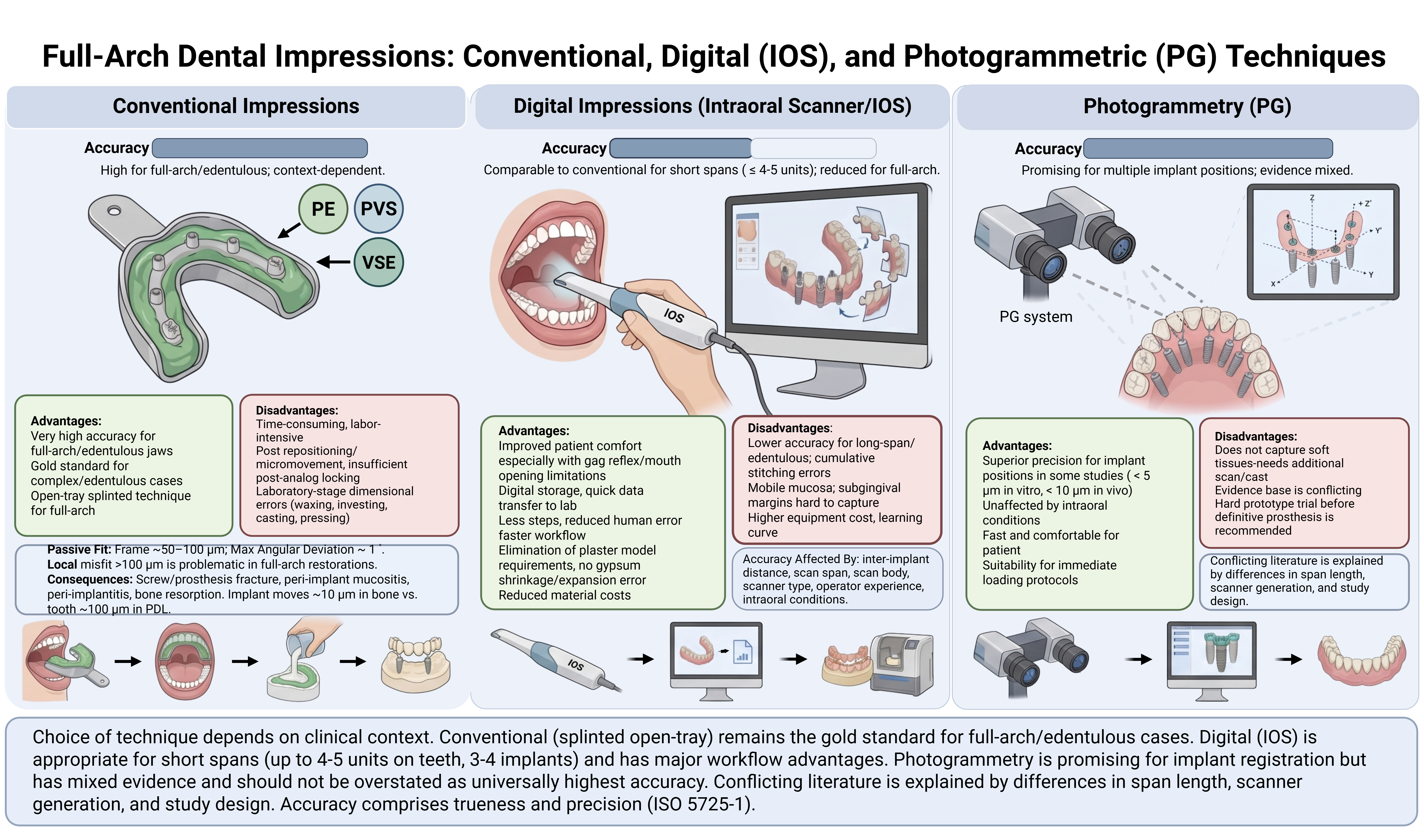
Advances in digital dentistry now permit the fabrication of full-arch prosthetic restorations through a fully digital workflow, extending from impression acquisition to the production of the definitive prosthesis. Intraoral scanning has become an acceptable alternative to conventional impression techniques, providing valuable information for diagnosis, restorative dentistry, orthodontic analysis, and implant-supported prosthodontics. Despite the many advantages offered by digital methods, conventional impression techniques using polyether, polyvinyl siloxane, and vinyl siloxanether materials remain widely used by clinicians because of their excellent dimensional stability and well-documented precision. This review compares and critically discusses conventional and digital impression techniques for acquiring full-arch dental registrations, with particular attention to their accuracy, advantages, limitations, and clinical indications. The accuracy of digital impressions is strongly influenced by inter-implant distance, scan span length, scan body geometry and material, scanner type, operator experience, and intraoral conditions such as mobile mucosa, saliva, and limited anatomical landmarks. For single-unit and short-span restorations of up to four or five units, intraoral scanning achieves accuracy comparable to that of conventional techniques. In long-span and full-arch restorations, however, accuracy tends to decline owing to cumulative image-stitching errors and the scarcity of reference points, a problem that is especially pronounced in completely edentulous arches. Photogrammetry has emerged as a promising alternative for the reliable registration of multiple implant positions, although it inadequately captures peri-implant soft tissues. Overall, technique selection depends primarily on the number of teeth or implants and the distance between them; conventional methods remain preferable for completely edentulous patients, whereas digital workflows are suitable for restorations spanning up to three or four implants. Despite persisting limitations, digital techniques are strong candidates to eventually outperform conventional approaches in dentistry.Full-arch impression, conventional impression, digital impression, intraoral scanner, photogrammetry, implant-supported prosthesis, impression accuracyEbru Sümer Ekin27.08.2026e260826 -
Can digital impression methods eliminate the current problems of conventional impression techniques?
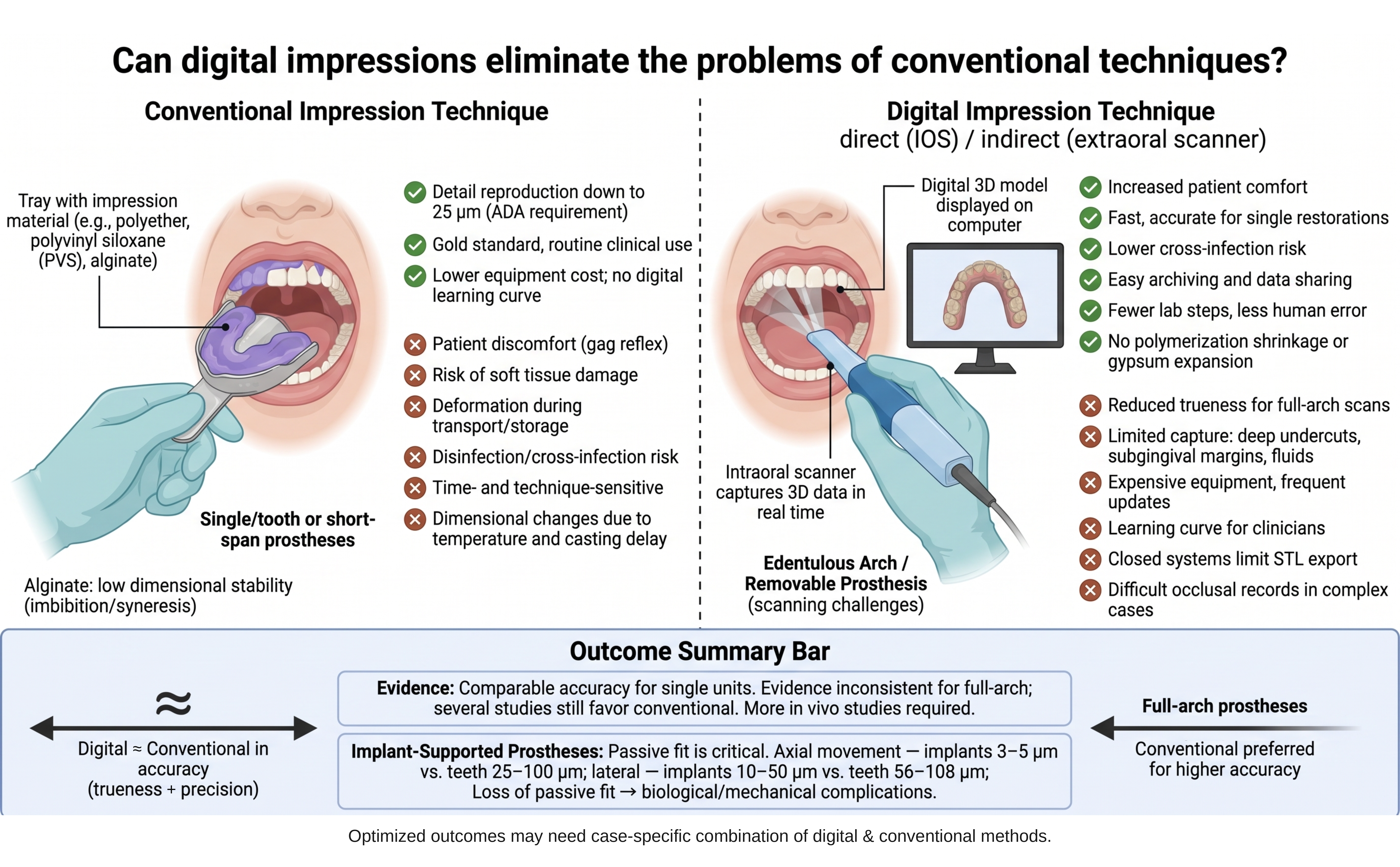
Conventional impression materials remain a cornerstone of routine dental practice, yet they carry well-documented drawbacks, including a pronounced gag reflex, limited patient comfort, potential soft-tissue trauma, dimensional changes with temperature and elapsed time, the need for disinfection, cross-infection risk, and susceptibility to distortion during transport, storage, and laboratory handling. Digital impression systems have been introduced to overcome these limitations, and their use in dentistry—particularly in prosthodontics—continues to expand. This review compares conventional and digital impression techniques and examines whether digital workflows can resolve the shortcomings inherent to conventional methods. Conventional elastomers such as polyether and addition-type silicones offer excellent dimensional stability and detail reproduction and are currently regarded as the gold standard. Digital impressions, obtained with intraoral or extraoral scanners, provide clear advantages in patient comfort, procedure time, reduced cross-infection risk, elimination of polymerization shrinkage and gypsum expansion, and convenient digital archiving and transfer. However, their accuracy is influenced by scanning area, scanner sensitivity, operator experience, moisture, patient movement, and inter-implant distance. The reviewed literature indicates that intraoral scanners achieve clinically acceptable accuracy for single units and single-quadrant restorations, whereas evidence for full-arch and multiple implant-supported prostheses remains inconsistent, with several studies still favoring conventional impressions. Accuracy—comprising trueness and precision—is especially critical for passive fit in implant-supported restorations. Most comparative data derive from in vitro models that do not reproduce intraoral conditions such as saliva, blood, and patient movement, underscoring the need for further in vivo research. In conclusion, although the benefits of digital impressions are evident, their long-term clinical efficacy and safety require additional investigation, and technique selection should weigh accuracy, time efficiency, learning curve, and cost. A case-specific combination of digital and conventional approaches may offer added benefit.Digital impression, intraoral scanner, conventional impression, trueness and precision, prosthodonticsYakup Kantaci27.08.2026e260815 -
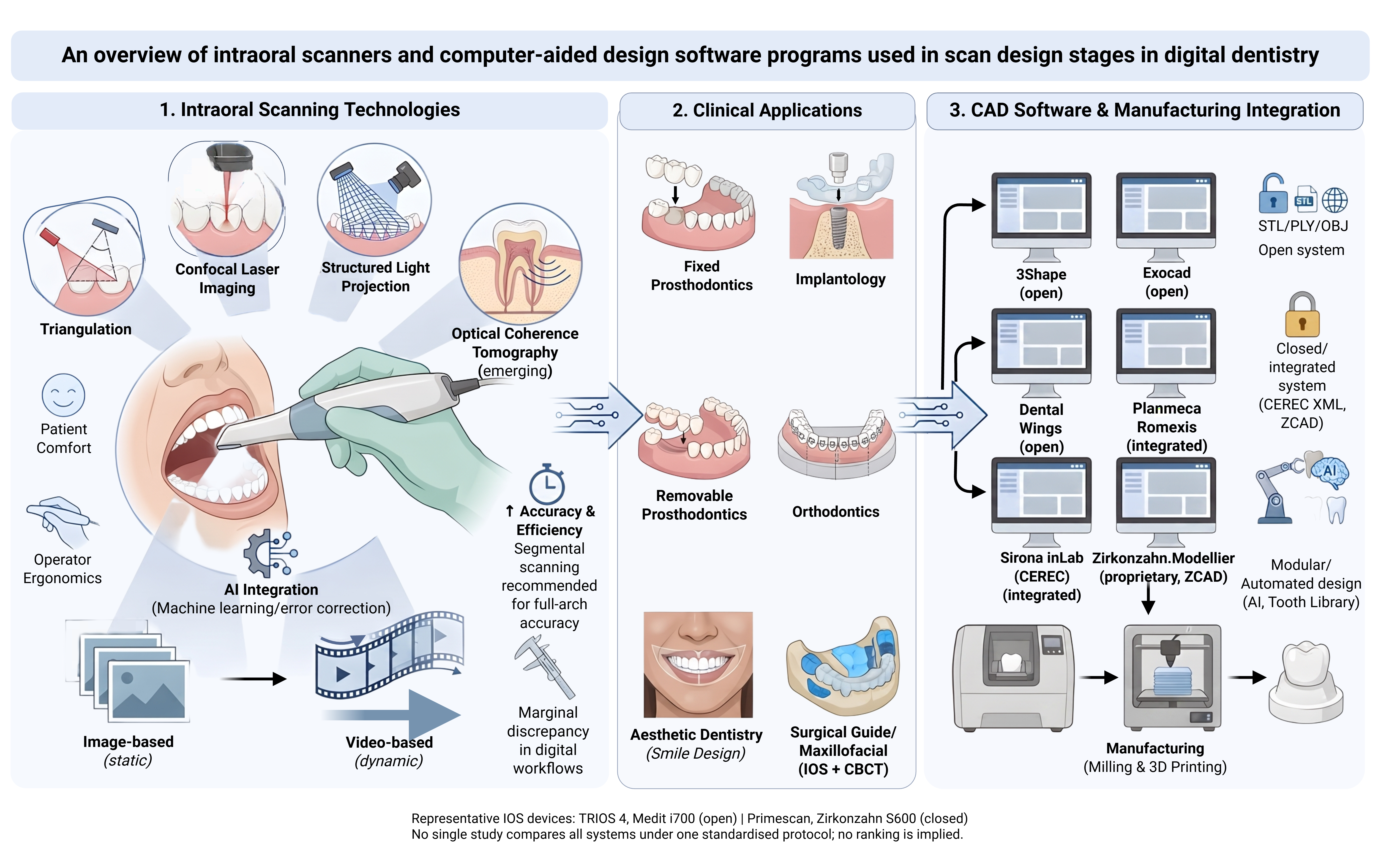
An overview of intraoral scanners and computer-aided design software programs used in scan design stages in digital dentistry
In prosthetic dental treatments, obtaining the negative of the prepared area in the most accurate way directly affects the success of the restoration to be made. Therefore, it is necessary to give importance to the impression material and the impression technique used. Impression materials have undergone changes over the years in order to record the most accurate and sensitive detail. Studies have tried to improve many physical and chemical properties of the impression material. Despite this, it should not be forgotten that there are numerous external factors such as the skill of the clinician, environmental conditions, and storage conditions for the impression to be obtained. Developments in computer-aided design and design (CAD/CAM) technology have significantly increased the use of these systems in dentistry in recent years. In dentistry, many companies in the market aim to minimize the problems that may occur in the laboratory and clinical environment by adding intraoral or extraoral scanners to CAD/CAM systems. The first step of dental CAD-CAM systems is digital scanning with an intraoral or laboratory scanner. This step is followed by the construction phase of restorations with digital design and production devices on the computer. Therefore, the success of these restorations depends largely on the accuracy and precision of the scanning devices. This review will discuss CAD/CAM systems and intraoral scanners and software programs.Digital dentistry, intraoral scanners, CAD/CAM systems, digital impression, scanning accuracy, digital workflowMuhammed Burak Süzen, Hayriye Yasemin Yay Kuşçu27.08.2026e260828 -
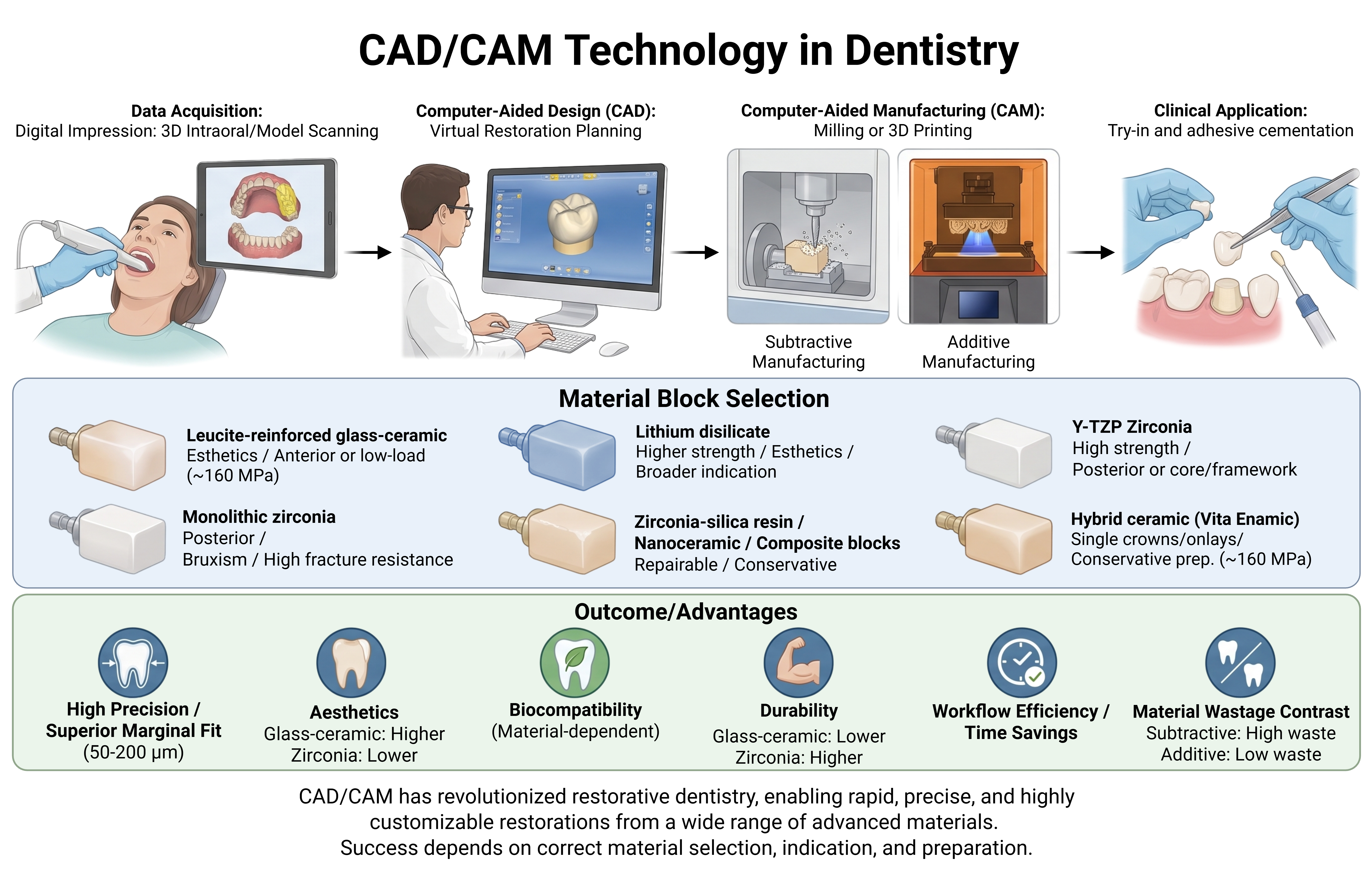
CAD/CAM technology in dentistry
The remarkable advancement of dental materials, treatment techniques, and prosthesis manufacturing processes has elevated computer-aided design and manufacturing (CAD/CAM) systems to a new level of clinical capability. Since its introduction to dentistry in the 1980s—beginning with the chairside CEREC system and the Procera framework concept—CAD/CAM has reshaped contemporary practice by enabling aesthetic, durable, and reliably reproducible restorations while transforming the way laboratories design and fabricate both framework (substructure) and monolithic restorations. Because the production stages are tightly controlled and the material blocks are industrially processed, modern CAD/CAM materials also offer improved structural properties—reduced microporosity, greater homogeneity, and lower firing shrinkage—relative to conventionally processed ones, while reducing laboratory steps, working time, and the overall error rate. This narrative review examines the principal CAD/CAM restorative materials, the digital data-processing workflow, and restoration production methods, together with their influence on marginal fit. The main material classes are described—leucite- and lithium-disilicate-reinforced glass ceramics, yttria-stabilized tetragonal zirconia polycrystals (Y-TZP) and monolithic zirconia, resin-matrix and nanoceramic blocks, composite blocks, and hybrid ceramics such as Vita Enamic—with their compositions, mechanical properties, indications, and limitations, including flexural strengths ranging from approximately 160 MPa for glass ceramics to substantially higher values for zirconia. The data-processing stage, comprising direct intraoral and indirect model scanning followed by computer-aided design of a virtual model, is reviewed, along with the two production routes: subtractive milling from prefabricated blocks and additive fabrication by selective laser sintering and 3D printing, which minimizes material waste. Marginal adaptation is discussed as a key determinant of restorative success, with reported clinically tolerable misfit values of 50 to 200 µm and CAD/CAM milling shown to provide superior marginal conformance to conventional techniques. Overall, CAD/CAM workflows offer measurable gains in material quality, accuracy, and efficiency over conventional methods, although optimal outcomes depend on correct material selection, appropriate indication, and proper tooth preparation.CAD/CAM, dental ceramics, monolithic zirconia, intraoral scanner, marginal adaptation, digital workflowZuhal Görüş, Hayriye Yasemin Yay Kuşçu, Nur Sena Alioğlu Koparal29.08.2026e260817
1 - 6 of 6 items





















 Open Access articles are distributed under the terms of the