
 10.5577/jomdi.e230310
Vol. 7 No. S1 (2026) Continuous Publication
View Current Issue
DENTISTRY
40 Items
All Items
-
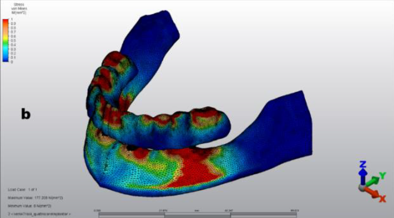
Does a sloped neck implant design facilitate All-on-4 surgeries? A three-dimensional finite element analysis
Aim: In the treatment of edentulous resorbed mandibles using All-on-4 concept, the distal part of posteriorly tilted implants with a conventional flat neck design is usually embedded in the bone, which may lead to improper installation or positioning of angled multiunit abutments. Bone trimming from the distal area, where the implant neck is embedded in the bone, is typically necessary to avoid this complication. This study aimed to evaluate the biomechanical behavior of a sloped marginal neck configuration of implant posteriorly tilted at 30° in edentulous mandibles using a three-dimensional finite element analysis (FEA). Methodology: The sloped neck implants were posteriorly tilted at 30° without any further operation in the edentulous mandibular model designed using the FEA. The tilted conventional flat neck implants were placed at the same length, diameter, and angle, and 2 mm 2 of bone was trimmed from the distal area. Similar multiunit abutments, two conventional flat neck implants axially placed on the anterior side, titanium frameworks, and acrylic resin prosthetic dentures were created for both models. The biomechanical behaviors of the models were evaluated by applying a foodstuff force of 50 N to the incisors, 100 N to the premolars, and 150 N to the molar cantilever. Results: The von Mises stress value of the sloped neck implants (158.489 MPa) was lower than that of the conventional flat neck implants (177.208 MPa). Both the maximum and minimum principal stress values in the surrounding cortical and cancellous bones were within the acceptable physiological limits. Conclusion: Sloped neck implants can be placed easily and are less invasive than conventional flat neck implants; thus, they can be a treatment alternative that can facilitate the All-on-4 concept. How to cite this article: Toprak ME. Does a sloped neck implant design facilitate All-on-4 surgeries? A three-dimensional finite element analysis. J Med Dent Invest 2023;4:e230310. https://doi.org/10.5577/jomdi.e230310 Linguistic Revision: The English in this manuscript has been checked by at least two professional editors, both native speakers of English.Dental implant, sloped neck design, bone resorption, biomechanics, finite element analysis, All-on-4Mehmet Emin Toprake230310 -

Use of platelet-rich fibrin (PRF) in the periodontology: A review
Platelets play an essential role in wound healing and periodontal regeneration, so using platelet concentrates can speed up wound healing after periodontal treatment. Platelet-rich fibrin (PRF), an autogenous concentrated blood product, is a fibrin matrix comprising molecular and cellular elements that allow for optimum improvement and PRF also increases the production of osteoprotegerin, which causes osteoblast proliferation and acts as an osteoconductive and osteoinductive material, initiating bone regeneration. Platelet-rich plasma (PRP) and PRF are platelet concentrates prepared from the patient's own blood. Recent studies have focused on the development of therapeutic alternatives that are simple to prepare, non-toxic or biocompatible to living tissues, and inexpensive, with the potential to result in the local release of growth factors that accelerate hard and soft tissue healing. PRP is a plasma fraction that contains a high concentration of growth factors. PRF is a natural fibrin-based biomaterial derived from an anticoagulant-free blood harvest without any artificial biochemical modification, allowing fibrin membranes enriched with platelets and growth factors to be obtained. It facilitates the application of growth factors to the surgical area in a concentrated form, thereby accelerating wound improvement and regeneration. Platelet concentrates are used in many areas of dentistry, including soft tissue improvement, plastic periodontal surgery, gingival enlargement, ridge preservation, regeneration of bone defects, drug-related osteonecrosis of the jawbone, sinus augmentation, immediate implant placement, and implant osseointegration. Their use is preferred due to their ease of application and low cost. This paper reviews the current literature concerning the utilization of platelet-rich fibrin in periodontology. How to cite this article: Sağsöz A, Acun Kaya F. Use of platelet-rich fibrin (PRF) in periodontology: A review. J Med Dent Invest 2023;4:e230317. https://doi.org/10.5577/jomdi.e230317 Linguistic Revision: The English in this manuscript has been checked by at least two professional editors, both native speakers of English.Platelet-rich fibrin, platelet-rich plasma, regeneration, autogenous blood product, growth factorsAslı Sağsöz, Filiz Acun Kayae230317 -

Evaluation of dentists’ approach to retreatment in Turkey: A survey study
Aim: This study aimed to evaluate the approaches that dentists working in Turkey take to retreatment. Methods: A questionnaire with 16 questions regarding gender, institution, specialty, experience, and root canal treatment stages was distributed to 223 dentists. The collected questionnaires were analyzed in R 4.0.0 programming language. A chi-square test analyzed the categorical variables. The statistical analyses were performed at a 5% significance level. Results: A statistically significant relationship was found between the years spent practicing dentistry and the methods of determining the length of root canal treatment ( p < 0.05). The relationship between the choices of irrigation agents for canal treatment and the professional title was found to be statistically significant ( p < 0.05). No significant relationship was found between gender and other parameters ( p > 0.05). A statistically significant difference was found between the treatment protocol applied in molar teeth with lesions, the success rate of the patients who underwent retreatment, and the practitioners’ specializations ( p < 0.05). The relationship between retreatment in unsuccessful root canal treatments and the institution studied was also statistically significant ( p < 0.05). A total of 66% of dentists working in Oral and Dental Health Center, 88% of dentists working in private clinics, and 90% of dentists working at universities reported that they performed retreatment. The relationships among the frequency of endodontic treatment, the use of magnification while performing root canal treatment, the group of teeth that was most often treated, and the institutions in which dentists work were also found to be statistically significant. Conclusion: In this survey study, which was conducted to evaluate the practice of endodontic treatment, the rate of dentists performing endodontic treatments was found to have increased. Endodontists performed retreatment cases at a higher rate than general practitioners. Dentists with less professional experience had higher rates of endodontic treatment for difficult–to–treat molars than those with more professional experience. How to cite this article: Tellioğlu Avcı GR. Evaluation of dentists’ approach to retreatment in Turkey: A survey study. J Med Dent Invest 2023;4:e230314. https://doi.org/10.5577/jomdi.e230324 Linguistic Revision: The English in this manuscript has been checked by at least two professional editors, both native speakers of English.Retreatment, endodontics, endodontics practice, surveyGüliz Rana Tellioğlu Avcıe230324 -

Effect of hesperidin on deltamethrin toxicity in rat periodontal membrane
Aim: Deltamethrin (DM), a hydrophobic and lipophilic substance used for insect control, easily accumulates in the body, increasing ROS production and causing mucosal inflammation and damage. The aim of this study was to explore the prophylactic effects of hesperidin on DM toxicity in periodontal membrane. Methods: The study was conducted for 14 days on 21 male Wistar albino rats. The experimental animals were divided into three groups (n=7): The Sham group received 0.9% NaCl (1 mL). The deltamethrin group received 0.5 mg/kg DM (1 mL, dissolved in dimethyl sulfoxide). The deltamethrin + hesperidin group received DM (0.5 mg/kg) + hesperidin (100 mg/kg) (both drugs dissolved in 1 mL dimethyl sulfoxide). All treatments were administered daily via the orogastric route for 14 days. At the end of the experiment, the rats were euthanized by cervical dislocation, and biochemical and histological analyses were performed. Results: The biochemical tests showed significantly higher malondialdehyde (MDA) levels in the deltamethrin group than in the Sham group. Combined application of hesperidin and deltamethrin resulted in significantly lower MDA levels in the deltamethrin + hesperidin group than in the deltamethrin group. Histopathological examination of the periodontal tissues of the deltamethrin group revealed cell degeneration at the alveolar bone-tooth junction, including atypical cells, irregularity in collagen fibers, dilatation and congestion of the blood vessels, and inflammatory cell infiltrates distributed in aggregate and solitary forms. The deltamethrin + hesperidin group showed substantially less cell degeneration. Conclusion: Hesperidin application helped alleviate deltamethrin toxicity by preventing oxidative stress and reducing cell degeneration. How to cite this article: İpek F, Deveci E, İpek P. Effect of hesperidin on deltamethrin toxicity in rat periodontal membrane. J Med Dent Invest 2023;4:e230332. https://doi.org/10.5577/jomdi.e230332 Linguistic Revision: The English in this manuscript has been checked by at least two professional editors, both native speakers of English.Hesperidin, deltamethrin, periodontal membrane, ratFikret İpek, Engin Deveci, Polat İpeke230332 -

Investigation of the prevalence of potentially malignant and malignant oral lesions in the Southeastern Anatolia Region of Turkey: A 5-year retrospective study
Aim: The aim of this study was to determine the region, frequency, distribution, age, and sex of histopathologically diagnosed oral malignant and oral potentially malignant lesions obtained from patients admitted to the Department of Pathology, Dicle University Faculty of Medicine, Southeastern Anatolia Region of Turkey. Methods: The pathological data of oral malignant and potentially malignant lesions histopathologically diagnosed at the Department of Pathology, Dicle University Faculty of Medicine, between 2018 and 2023, were retrospectively reviewed. Potentially malignant and malignant oral lesions were classified, and their distribution according to region, age, and sex was analyzed. The Shapiro-Wilk test was used to analyze the conformity of continuous variables to the normal distribution. The type I error level was accepted as 5% in statistical analyses. Results: Among 161 patients with oral or potential malignancy, 76 (47.2%) were female and 85 (52.8%) were male, with a mean age of 53.9 ± 17.4 years. The most common lesions were found in the buccal mucosa (40.4%). When 104 malignant lesions were analyzed, the most common lesion was oral squamous cell carcinoma with a rate of 75%, and when 57 potentially malignant lesions were analyzed, 38 (66.7%) of the patients had lichen planus, and 19 (33.3%) had leukoplakia. Conclusion: This retrospective study shows the distribution, frequency, and characteristics of malignant lesions in the oral cavity in a group of populations, especially in the Southeastern Anatolia Region of Turkey, and allows comparison with studies in different populations. How to cite this article: Kaplan B, Alabalık U, Akşahin C, Çolak M, Ortaç H. Investigation of the prevalence of potentially malignant and malignant oral lesions in the Southeastern Anatolia Region of Turkey: A 5-year retrospective study. J Med Dent Invest 2023; 4: e230348. https://doi.org/10.5577/jomdi.e230348Malignant oral lesion, potentially malignant oral lesion, oral squamous cell carcinoma, histopathology, retrospective studyBahar Kaplan, Ulaş Alabalık, Cihat Akşahin, Mehmet Çolak, Hatice Ortaçe230348 -

Demographic investigation of 700 cases with severe odontogenic abscess in the maxillofacial region: A 6-year retrospective study
Aim: The aim of this retrospective study was to retrospectively analyze the demographic distribution and treatment methods of patients with odontogenic abscess who applied to Dicle University, Faculty of Dentistry, Department of Oral and Maxillofacial Surgery between 2017 and 2022, and evaluate the findings. Methods: Patient documents of 700 odontogenic abscess cases who applied to the Dicle University, Faculty of Dentistry, Department of Oral and Maxillofacial Surgery clinic were collected and examined. The data obtained were analyzed according to gender, age, time, anatomical localization, and treatment options. All data obtained were evaluated with statistical analysis. Results: As a result of the statistical analysis; a total of 700 patients, including 400 men (57%), and 300 women (43%), were treated. Of the patients with a mean age of 33±18 years, 63% (440) were treated with extraoral drainage and 37% (260) with intraoral drainage. In terms of localization, the most common areas of odontogenic abscess formation were mandibular posterior (54% - 376 cases), maxillary posterior (26% - 184 cases), maxillary anterior (17% - 116 cases), and mandibular anterior (3% - 24 cases). The most common reasons patients came to the clinic were swelling, pain, fever, and trismus. The year 2022 was analyzed as the year with the most intensive patient treatment, and 2017 as the year with the least patient treatment. In the analyses, when looking at the distribution of gender and localization by years, there was no statistically significant difference ( p > 0.05), but when looking at the distribution of drainage techniques by years, it was determined that there was a statistically significant difference ( p < 0.001). Conclusion: Demographic characteristics and treatment options of patients with odontogenic abscesses vary depending on the causative tooth area. Odontogenic infections can become life-threatening in a short time if left untreated. Intraoral and extraoral drainage are very important for success in the treatment of common odontogenic abscesses where medical treatment is inadequate. How to cite this article: Güler R. Demographic investigation of 700 cases with severe odontogenic abscess in the maxillofacial region: A 6-year retrospective study. J Med Dent Invest 2023; 4: e230350. https://doi.org/10.5577/jomdi.e230350Maxillofacial infection, odontogenic infection, abscess, drainageRıdvan Gülere230350 -

Effect of different polishing methods applied to denture base materials on microbial adhesion and surface roughness
Aim: The aim of our study was to investigate the effect of various polishing processes applied to different types of PMMA (polymethyl methacrylate) denture base materials on the surface roughness and microbial adhesion of the base material. Methods: Sixty specimens were prepared from heat-polymerized conventional PMMA (G), injectable PMMA (E), and pre-polymerized CAD-CAM PMMA (C) denture base materials. All the sample surfaces were sanded with silicon carbide abrasives of 800, 1000, 1200, and 2500 grit. The specimens of each denture base material were divided into three subgroups (n=20), in which different polishing methods were applied. The first group was mechanically polished (M), and the other two groups were chemically polished with light-polymerized glaze liquids [Palaseal (Ps), Optiglaze Color (Og)]. A profilometer was used to evaluate the surface roughness. Colony counting was performed to assess bacterial colonization after one day. The data obtained from all these measurements were evaluated using Analysis of Variance and Bonferroni Corrected test was used for multiple comparisons of means ( p = 0.05). The interaction between surface roughness and bacterial colonization was examined using the Pearson’s correlation test. Regression analysis was performed to determine the relationships between them. Results: When PMMA base materials were compared, the lowest average surface roughness (Ra) and bacterial colonization values were obtained in Group C. When the polishing methods were compared, the lowest average surface roughness values were obtained for Group M. As a result of the bacterial colonization measurement, lower colonization values were obtained with standardized mechanical polishing compared to the other groups. Conclusion: There was no difference between the different PMMA base materials in terms of surface roughness; however, there were statistically significant differences in bacterial colonization. Our study showed that mechanical polishing yielded better results than light-activated glazing agents. How to cite this article: Yarar S, Değer Y, Tekin S. Effect of different polishing methods applied to denture base materials on microbial adhesion and surface roughness. J Med Dent Invest 2023; 4: e230351. https://doi.org/10.5577/jomdi.e230351CAD-CAM, digital, PMMA, polishing, surface roughnessSadi Yarar, Yalçın Değer, Samet Tekine230351 -

Impact of malocclusion on Oral Health-Related Quality of Life (OHRQoL) using the Oral Health Impact Profile (OHIP-14)
Aim: The present study aims to examine the oral health-related quality of life (OHRQoL) using the Oral Health Impact Profile (OHIP-14) among patients with orthodontic complaints in terms of malocclusion classification from the aspects of gender and age. Methods: The present study was carried out using face-to-face interviews with 150 patients (73 females and 77 males) aged between 12 and 34 years. Demographic information, including age and gender, was requested from the patients via the questionnaire form. Participants were requested to complete the abbreviated version of the OHIP-14 to assess the OHRQoL. Patients were classified into three groups: Class I (n = 50), II (n = 65), and III (n = 35) by Angle classification. In order to measure the OHRQoL, the patients were asked to answer the Turkish version of the Oral Health Impact Profile (OHIP-14). The statistical analyses were performed using ANOVA, Bonferroni, and the Student’s t-test. The statistical significance level was set at p < 0.05. Results: In OHRQoL, the Class III group was found to have statistically significantly higher values when compared to the Class II and Class I groups ( p < 0.05). The mean scores in the psychological discomfort and handicap subscales were found to be statistically significantly higher ( p < 0.05). Conclusion: Malocclusion has a negative effect on OHRQoL. The increasing severity of malocclusion causes lower OHRQoL. How to cite this article: Topal R. Impact of malocclusion on Oral Health-Related Quality of Life (OHRQoL) using the Oral Health Impact Profile (OHIP-14). J Med Dent Invest 2023; 4: e230354. https://doi.org/10.5577/jomdi.e230354Malocclusion, oral health-related quality of life, Oral Health Impact Profile-14, orthodontic patient, orthodontic treatmentRefika Topale230354 -

Finite element stress analysis in mandibular molars with apical root resorption and crown destruction after root canal treatment using various materials
Aim: The objective of this study was to analyze the stress distribution of root canal treated teeth under occlusal forces and aid in the selection of appropriate materials for teeth with root tip resorption and crown destruction, using the finite element stress analysis method (FEM). Methods: The study was conducted on two-rooted lower molar teeth, divided into five groups. The root length was 14 mm, the crown length was 7.5 mm, and the total length was 21.5 mm. It was assumed that the distal canals were resorbed by 4 mm. Group 1 consisted of untreated intact teeth. Group 2 involved filling the mesial canals with gutta-percha, filling the apical part of the distal canal with 4 mm Mineral Trioxide Aggregate (MTA), and fabricating the crown using glass fiber post and IPS e.max materials. Group 3 involved filling the mesial canals with gutta-percha, filling the apical part of the distal canal with 4 mm Biodentin, and manufacturing the crown using glass fiber post and IPS e.max materials. Group 4: The canals were filled with MTA, and an IPS e.max endocrown was used. Group 5: The canals were filled with Biodentin and an IPS e.max endocrown was used. The study examined the effect of a 100 Newton force applied in the occlusal direction on dental tissues using finite element stress analysis. Results: The study found that the application of fiber posts resulted in less stress accumulation in tooth tissues compared to endocrown. Conclusion: The use of Biodentin or MTA in endocrown teeth did not affect the stress in tooth tissues caused by applied force. In conclusion, while fiber posts increase durability by reducing stress in root-end resorbed teeth, the effect of Biodentin or MTA on stress resistance in dental tissues is limited. How to cite this article: Duran S, Şirin DA, Topbaş C. Finite element stress analysis in mandibular molars with apical root resorption and crown destruction after root canal treatment using various materials. J Med Dent Invest 2024;5:e240320. https://doi.org/10.5577/jomdi.e240320Apical Resorption, Biodentine, endocrown, finite element analysis, glass fiber post, mineral trioxide aggregateSerkan Duran, Dursun Ali Şirin, Celalettin Topbaşe240320 -

Clinical applications and improvement of resin bonding for PEEK in resin-bonded fixed dental prosthesis: A review
Resin-bonded fixed dental prostheses (RBFDPs) are a commonly used precision treatment, such as fixed dental prosthesis (FDP) and implant-supported dentures. Full crown preparation of vital teeth, which is required for traditional FDPs, may result in a risk of pulp reaction as a large portion of the coronal tooth structure must be removed. RBFDP has been recognized as a non-invasive or minimally invasive treatment modality for the replacement of single missing teeth. RBFDPs require much less tooth preparation than conventional FDPs. These dental treatments are restorative solutions used to cover missing teeth and achieve an aesthetic smile. The biomechanical rules, aesthetics, polishability, and bond strength have been emphasized in RBFDP treatments. Polyetheretherketone (PEEK) is one of the most widely used biomaterials in dentistry. The material, which stands out especially with its acceptable aesthetics, biocompatibility and modulus of elasticity close to dentin, has enabled it to be used in RBFDP treatments. Since PEEK is lighter than metal alloys, patients experience less discomfort and a feeling closer to natural teeth. Furthermore, the thermoplastic properties of PEEK are claimed to provide a precise fit, allowing the restoration to better integrate into the natural tooth structure. In addition, its ability to be customized using different modern manufacturing techniques is thought to offer dentists and patients a reliable option for successful long-term treatment. Our review is about the use of PEEK material as an alternative material in RBFDPs with low negative effects on periodontal tissues and their bonding with resins.Fixed dental prostheses, PEEK, Polyetheretherketone, polymer, resin-bondedBahar Tekin, Ayşe Rençber Kızılkaya, Vahti Kılıç, Metin Bakır, Samet Tekin, Suzan Cangüle240325 -

Surface characterization and optical properties of zirconia-reinforced lithium silicate ceramics against different mouthwashes and thermocycling
Aim: To evaluate color stability (ΔE 00 ), translucency (TP), and surface roughness (Ra) of zirconia-reinforced lithium silicate (ZLS) with lithium disilicate (LS) against different mouth rinses after thermocycling. Methods: A total of 270 specimens were prepared: LS (IPS Emax CAD (group EC)), ZLS-1 (Vita Suprinity (group VS)), and ZLS-2 (Celtra Duo (group CD)). For each specimen, ΔE 00 , TP, and Ra measurements were recorded with a clinical spectrophotometer (CM-3600A) and a 3D non-contact profilometer (AEP Nanomap-1000WLI), respectively, between baseline and after 10000 cycles of thermal cycling. Each group was divided into six subgroups (n=15) according to the immersion solution: five mouthwashes (Meridol, Klorhex, Listerine Cool Mint, Listerine Healthy White Natural Lemon&Salt, and Curasept ADS 212), and distilled water for 72 hours. Statistical analysis was performed by using a three-way robust ANOVA with Tukey HSD test (α=0.05). Results: Listerine Lemon&Salt and Cool Mint caused the highest ΔE 00 and Ra values, while TP values of tested mouthwashes were similar ( p =0.051). Group EC had the highest ΔE 00 and Ra values with the lowest TP values against mouthwashes ( p <0.05). Conclusion: Prolonged use of mouthwashes may cause color change and increased surface roughness in LS more than ZLS. Patients should be advised to use alcohol-containing mouthwashes with caution in daily use.Mouthwashes, lithium disilicate, zirconium oxide, Vita Suprinity, MeridolMerve Benli, Müge Yargıcı Çağlayan, Beril Eker Gümüşe240334 -

Effects of two different chelation solutions and activation protocols on the push-out bond strength of a bioceramic sealer
Introduction: This study evaluated the effects of glycolic acid (GA) and ethylenediaminetetraacetic acid (EDTA) chelation solutions on the push-out bond strength of AH Plus Bioceramic Sealer when activated using passive ultrasonic irrigation (PUI) and XP-endo Finisher (XPF) activation protocols. Materials and Methods: A total of 48 similarly sized extracted mandibular premolars were randomly assigned to four groups (n = 12). All canals were prepared using Reciproc (25.08) instruments. Irrigation was performed using 5 ml of 5% sodium hypochlorite (NaOCl) between instruments, totaling 20 ml of NaOCl per canal. The teeth were divided into four groups for the final irrigation: Group 1: GA+PUI; Group 2: GA+XPF; Group 3: EDTA+PUI; and Group 4: EDTA+XPF. NaOCl was activated for 1 minute in all groups, followed by chelator activation for 30 seconds with the designated activation system. Canals were filled using the single-cone technique and AH Plus Bioceramic Sealer. The samples were incubated for one week. A single horizontal middle dentin section of 2 ± 0.1 mm thickness was prepared from each root. Push-out bond strength was tested using a universal testing machine with a loading rate of 0.5 mm/min. The data were analyzed using the Kruskal–Wallis H and Mann–Whitney U tests, with a p < 0.05 significance level. Results: There were no statistically significant differences among the GA+PUI, GA+XPF, EDTA+PUI, and EDTA+XPF groups (p > 0.05). Conclusion: Both EDTA and GA showed similar bond strength effectiveness when activated by either irrigation activation system. How to cite this article: Batur M, Kılıvan H, Yeniçeri Özata M, Tellioğlu Avcı GR. Effects of two different chelation solutions and activation protocols on the push-out bond strength of a bioceramic sealer. J Med Dent Invest 2024;5:e240342. https://doi.org/10.5577/jomdi.e240342Passive ultrasonic irrigation, XP-endo Finisher, push-out bond strength, EDTA, glycolic acidMuharrem Batur, Hasret Kılıvan, Merve Yeniçeri Özata, Güliz Rana Tellioğlu Avcıe240342 -

Comparison of cyclic fatigue resistance of different rotational and reciprocal file systems
Aim: The purpose of this study is to compare the cyclic fatigue resistance of the TruNatomy and ProTaper Next file systems, which work rotational movements, and Reciproc and WaveOne Gold file systems, which work reciprocal movements. Methods: ProTaper Next X2 (25/.06), WaveOne Gold Primary (25/.07), Reciproc R25 (25/.08), and TruNatomy Prime (26/.04) files were used in the cyclic fatigue test. Four groups, 15 files from each group and a total of 60 files, were rotated in an artificial canal made of curved stainless steel with a curvature angle of 60 degrees, an inner diameter of 1.5 mm, and a radius of curvature of 3 mm. The breaking times of the files were determined with a digital stopwatch, and this value was converted into the number of cycles until breaking. Compliance with normal distribution was examined with the Shapiro‒Wilk test. The number of cycles until fracture for the respective files, which did not follow a normal distribution, were compared using the Kruskal‒Wallis test and multiple comparisons were made with the Dunn test. The analysis results were presented as mean ± standard deviation and median (minimum-maximum). The significance level was taken as p <0.050. Results: While the TruNatomy system had the highest number of cycles until fracture occurred, no statistically significant difference was found between the Reciproc and WaveOne Gold systems. The ProTaper Next system showed the lowest cyclic fatigue resistance. Conclusion: Within the limitations of the present in vitro study, TruNatomy, which makes rotational motion, showed the best cyclic fatigue resistance. The ProTaper Next system was the file with the lowest cyclic fatigue resistance. Obtaining similar results for files produced in different shapes and with different movement kinematics allowed us to conclude that cyclic fatigue is multifactorial. How to cite this article: Akın Tartuk G, Kaya S, Al Tai TN. Comparison of cyclic fatigue resistance of different rotational and reciprocal file systems. J Med Dent Invest 2024;5:e240347. https://doi.org/10.5577/jomdi.e240347Cyclic fatigue, endodontic file, ProTaper Next, Reciproc, TruNatomy, WaveOne GoldGizem Akın Tartuk, Sadullah Kaya, Tamara Nabhan Al Taie240347 -

Root canal configuration in mandibular anterior teeth: Insights from cone-beam computed tomography in a Turkish subpopulation
Aim: The aim of this study was to examine the structures, roots and canal numbers of mandibular anterior teeth using cone-beam computed tomography (CBCT). Methods: The study evaluated 4830 fully developed mandibular anterior teeth from 953 CBCT scans. The final sample consisted of 515 scans of females and 438 scans from males, with ages ranging from 17 to 69 years. Among the assessed teeth, 2475 were located on the right side and 2355 on the left, including 2614 from females and 2216 from men. The teeth were free of any fillings, resorption, or cracks in the root or crown. The study analyzed the numbers of roots and root canals, location of the apical foramen, and canal type according to Vertucci’s classification. Results: 82.7% of teeth had single-root canals, whereas four lateral teeth (0.2%) and 42 lower canines (2.4%) had two-roots. The apical foramen was located centrally in 80.9% of the teeth (n = 3909), followed by buccal (8.8%), distal (4.2%), lingual (3.4%), and mesial (2.7%) positions. Using Vertucci’s classification of root canal types, type-I was the most prevalent (65.3%), followed in order by types-III (28.6%), II (4.2%), V (1.5%), and VI (0.4%). Conclusion: Turkish mandibular anterior teeth have intricate anatomy. To enhance comprehension of the complex anatomy of Turkish mandibular incisors and canines, including the numbers of canals and roots and canal configurations, further studies of more patients from diverse regions of Türkiye are needed. The tooth configuration was influenced by gender rather than by tooth position. How to cite this article: Topbaş C, Atav A. Root canal configuration in mandibular anterior teeth: Insights from cone-beam computed tomography in a Turkish subpopulation. J Med Dent Invest 2024;5:e240362. https://doi.org/10.5577/jomdi.e240362Root canal morphology, Vertucci, apical foramen, cone-beam computed tomography, mandibular anterior teethCelalettin Topbaş, Ayfer Atave240362 -
Diagnosis and treatment methods in inferior alveolar and lingual nerve injuries: A review
Injuries to the inferior alveolar nerve (IAN) and lingual nerve (LN) are common complications of oral and maxillofacial surgery, frequently occurring during third molar extractions, implant placements, and orthognathic procedures. These injuries can lead to sensory deficits and negatively affect the patients’ quality of life. Although some cases resolve spontaneously, early diagnosis and appropriate treatment, including microsurgical repair, are often necessary to optimize the outcomes. This review aims to summarize the current approaches for the diagnosis and treatment of IAN and LN injuries and highlight emerging techniques in nerve regeneration. A comprehensive literature review was conducted to evaluate the classification of nerve injuries (Seddon and Sunderland), diagnostic modalities such as nerve stimulation tests and magnetic resonance neurography, timing of surgical intervention, and available treatment options, including neurorrhaphy, autografts, allografts, nerve conduits, pharmacologic therapies, and adjunctive regenerative strategies. Mild nerve injuries may recover spontaneously within 3–9 months; if sensory function does not return within this period, surgical repair is generally indicated. Early surgical intervention (within 3–6 months) yields more favorable outcomes, although later repair can still be beneficial. Direct neurorrhaphy is recommended for small nerve gaps (≤5 mm), whereas larger defects require grafting. Autografts, such as the sural or great auricular nerves, are the gold standard because of their compatibility; however, they entail donor-site morbidity. Processed allografts and conduits offer viable alternatives with reduced surgical time, although they can be costly and less predictable. Adjunctive therapies, including platelet-rich fibrin, corticosteroids, and low-level laser therapy, have demonstrated potential benefits but require further clinical validation. New biomaterials that incorporate stem cells and growth factors show promise for enhancing nerve regeneration. Effective management of IAN and LN injuries requires timely diagnosis and an individualized treatment plan based on the injury severity and patient factors. Early surgical repair improves the likelihood of functional recovery. While autografts remain the preferred method for reconstructing large nerve gaps, processed allografts and nerve conduits are valuable alternatives. Ongoing advances in regenerative materials and adjunctive therapies hold potential to improve outcomes and reduce complications associated with peripheral nerve repair.inferior alveolar nerve injury, lingual nerve repair, peripheral nerve regeneration, microsurgical neurorrhaphy, nerve grafting techniquesMehmet Sait Şimşek, İrfan Üstündağ, Sudenur Kocamemik1-7 -
Peri-implantitis and treatment: A review
The increasing use of implant therapy has also led to a higher prevalence of peri-implant diseases. Peri-implant diseases were defined at the 2017 World Workshop as biofilm-associated pathological conditions affecting dental implants and were classified as peri-implant mucositis and peri-implantitis. This review briefly addresses the etiopathogenesis, clinical features, and diagnosis of peri-implantitis, and discusses both nonsurgical approaches including the use of antiseptics and antibiotics, mechanical debridement, ozone therapy, probiotics, laser and photodynamic therapy, and platelet-rich fibrin (PRF) applications and surgical strategies such as resective and regenerative procedures. While short-term studies have reported favorable outcomes with surgical approaches, long-term follow-up has revealed various complications. Regenerative therapies, on the other hand, have demonstrated more promising results; therefore, careful soft tissue management and increasing the amount of keratinized gingiva and sulcus depth prior to surgical intervention are essential to enhance treatment success and maintain oral hygiene.dental Implants, peri-implantitis, guided tissue regenerationMusa Özdemir, Mehmet Sait Şimşek1-6 -
Endodontic treatment in primary teeth
Endodontic treatment in primary teeth, commonly referred to as pulpectomy, is an essential clinical procedure aimed at preserving primary teeth affected by pulp or periradicular infections until their physiological exfoliation. Maintaining primary teeth is crucial for proper mastication, speech development, arch integrity, and guidance of permanent tooth eruption. This comprehensive review summarizes current concepts, materials, and techniques used in the root canal treatment of primary teeth, with a focus on improving clinical outcomes. The complex and dynamic root canal anatomy of primary teeth, particularly the presence of physiological root resorption, presents unique challenges for endodontic procedures. Successful treatment depends on accurate diagnosis, careful case selection, effective chemomechanical preparation, optimal irrigation, and the use of appropriate root canal filling materials. Irrigation plays a critical role in eliminating microorganisms that cannot be removed by mechanical instrumentation alone. Commonly used irrigants include sodium hypochlorite, chlorhexidine, ethylenediaminetetraacetic acid, citric acid, and newer combination solutions, each with specific advantages and limitations regarding antimicrobial activity, tissue dissolution, and biocompatibility. Advances in instrumentation, particularly the use of nickel–titanium rotary systems specifically designed for pediatric patients, have improved cleaning efficiency, reduced treatment time, and enhanced patient cooperation. Determination of working length using electronic apex locators in combination with radiography has further increased procedural accuracy while minimizing radiation exposure. Root canal filling materials for primary teeth must meet specific criteria, including biocompatibility, adequate antimicrobial properties, resorption compatible with physiological root resorption, and safety for the underlying permanent tooth germ. Zinc oxide eugenol, calcium hydroxide–based pastes, iodoform-containing materials, and newer bioceramic-based pastes are commonly used, although no material fully satisfies all ideal requirements. In conclusion, endodontic treatment in primary teeth is a reliable and effective approach when performed with proper technique and material selection. Continuous updates in materials and technologies, along with adherence to evidence-based protocols, are essential for enhancing treatment success and supporting the overall oral health and development of pediatric patients.Primary tooth root canal treatment, root canal filling materials, irrigation solutions, chemomechanical preparationGizem Karagöz Doğan, Yelda Polat, Sema Çelenke250069 -
Non-fluoride systems for enamel remineralization: Types and mechanisms
Dental caries is a significant global public health issue, despite advancements in oral hygiene practices and the emergence of new protective substances. Although it is a progressive chronic condition, can be prevented and remineralized at an early stages without the formation of cavities. Minimally invasive dentistry emphasizes the importance of preventing strategies for remineralization of White spot lesions. New approaches are being developed to promote deeper lesion remineralization, minimize the risks associated with high fluoride levels, and ensure long-term carries control. Non-fluoride remineralization can be classified into two categories: those that enhance the efficacy of fluoride and biomimetic systems that imitate enamel regeneration. The aim of this brief paper is to review the some commonly used non-fluoride remineralization systems and mechanisms, challenges, and evidence underpinning some of the technological advances in enamel remineralization.Dental caries, remineralization, non-fluoride remineralization agentsBetül Kargül, Nur Kodaman Dokumacıgil, Ayşe Nur Parlakyıldız Gökçee250074 -
Use and misuse of antimicrobials in pediatric dentistry
The prescription of antimicrobials is a common practice in pediatric dentistry for the management of oral infections, trauma, and prophylactic indications. However, inappropriate use and misuse of these agents remain a significant concern, contributing to antimicrobial resistance, adverse drug reactions, and unnecessary exposure in children. The aim of this review was to evaluate the appropriate indications, clinical considerations, and common causes of misuse of antimicrobials in pediatric dental practice, with particular emphasis on antibiotics and analgesics. Children present unique anatomical, physiological, and behavioral characteristics that influence drug selection, dosage, and administration. Antibiotics are indicated primarily in cases of spreading odontogenic infections, facial cellulitis with systemic involvement, specific traumatic injuries, salivary gland infections of bacterial origin, and selected medically compromised conditions requiring prophylaxis. In contrast, antibiotics are not indicated for localized dental conditions such as irreversible pulpitis, necrotic pulps, localized apical abscesses without systemic signs, plaque-induced gingivitis, or viral infections, where definitive dental treatment and local measures are sufficient. Despite clear guidelines, antibiotic overuse in pediatric dentistry is frequently driven by misdiagnosis, time constraints, parental pressure, and insufficient knowledge of evidence-based indications. Such practices increase the risk of antibiotic resistance and may delay definitive dental care. The review also discusses the role of analgesics and anti-inflammatory drugs in pediatric dentistry, emphasizing that pain management should primarily rely on agents such as acetaminophen and ibuprofen rather than antibiotics. In conclusion, antimicrobials play an essential but limited role in pediatric dental care. Judicious prescribing based on accurate diagnosis, clinical severity, and established guidelines is critical to minimize misuse. Increasing practitioner awareness, improving education, and adhering to evidence-based protocols are key strategies to promote rational antimicrobial use and safeguard both individual and public health.Antimicrobial, misuse, overuse, antibiotics, analgesics, pediatric dentistryAyli̇n İslame250067 -
Pediatric dental approaches in children with chronic kidney disease
Chronic kidney disease (CKD) in childhood is a progressive and irreversible condition that affects multiple organ systems, including the oral and dental structures. This review article provides a comprehensive overview of the oral manifestations of CKD in pediatric patients and discusses appropriate dental management strategies. As the prevalence of CKD in children has increased in recent years, awareness of its oral implications has become increasingly important for pediatric dentists. Children with CKD commonly present with distinct oral findings resulting from metabolic disturbances, uremia, altered saliva composition, and long-term medical treatments. Although these patients often have inadequate oral hygiene and a carbohydrate-rich diet, several studies report a lower prevalence of dental caries compared to healthy peers. This paradox is mainly attributed to increased salivary urea levels, higher buffering capacity, and an alkaline oral environment, which inhibit acidogenic bacteria. However, the same salivary changes contribute to increased dental calculus formation. Additional oral findings include halitosis, gingival and periodontal alterations, oral mucosal pallor due to anemia, bleeding tendencies, and opportunistic infections, particularly in immunosuppressed or transplant patients. Renal osteodystrophy may affect the alveolar and supporting bone, increasing the risk of abnormal bone healing and jaw fractures following dental procedures. Moreover, medications commonly used in CKD management, such as cyclosporine and calcium channel blockers, may induce gingival overgrowth and xerostomia. Early identification of oral manifestations, regular dental follow-up, and preventive care are essential in children with CKD. A multidisciplinary approach involving pediatric dentists and pediatric nephrologists is crucial to ensure safe dental treatment and to improve both oral health outcomes and overall quality of life in this vulnerable population.Chronic kidney disease, nephropathy, oral manifestations, pediatric dentistry, renal failureÖzlem Beren Satılmış, Akif Demirel, Nurhan Özalpe250064 -

Comparative evaluation of the accuracy and completeness of responses provided by ChatGPT-5, Gemini 2.5 Flash, Grok 4, and DeepSeek-R1 to open-ended questions on cracked and fractured teeth
Aim: The aim of this study was to evaluate and compare the accuracy and completeness of responses to open-ended questions related to chronic cracked and fractured teeth given by four recently introduced large language models (LLMs): ChatGPT-5, Grok 4, Gemini 2.5 Flash, and DeepSeek-R1-0528. Methods: A total of 25 open-ended questions related to cracked and fractured teeth were prepared. The responses to these questions given by the LLMs were evaluated in terms of accuracy and completeness by restorative and endodontic specialists with 15 years of experience. Accuracy was evaluated using a 5-point Likert scale, and completeness was evaluated using a 3-point Likert scale. The Intraclass Correlation Coefficient (ICC), Shapiro-Wilk test, Kruskal-Wallis test, Dunn-Bonferroni test, and Spearman correlation analysis were used in the statistical analyses of the study. Results: The responses given to the questions about the diagnosis, causes, and treatment of cracked and fractured teeth showed significant differences among the four artificial intelligence (AI) models (p=0.015). In the subgroup analysis, the mean points of ChatGPT-5 (5±0) were significantly higher than those of DeepSeek-R1-0528 (4.48±0.77) (p=0.013). No significant difference was observed in the other paired comparisons (p>0.05). The completeness points also showed a difference between the models (p=0.028). The mean points of ChatGPT-5 (3±0) were significantly higher than those of DeepSeek-R1-0528 (2.76±0.44) (p=0.046), and no significant difference was observed in the other paired comparisons (p>0.05). Conclusion: Differences in terms of accuracy and completeness were observed between the LLMs in the responses given to open-ended questions about cracked and fractured teeth. The lowest mean points were obtained by DeepSeek-R1-0528, followed by Grok 4 and Gemini 2.5 Flash, with the highest points obtained by ChatGPT-5. It must not be forgotten that the results obtained can show variability according to the subject, the form of the question, and the content. Therefore, further studies encompassing both similar and different subjects are needed. It must also not be ignored that misinformation in healthcare services can lead to serious consequences.Artificial intelligence, large language models, ChatGPT, Gemini, Grok, DeepSeek, cracked tooth, fractured tooth, accuracy, completenessMakbule Taşyürek, Özkan Adıgüzel, Suzan Cangül, Hatice Ortaçe250161 -
Potential complications and preventive measures in pediatric dental treatments performed under general anesthesia and sedation
The aim of this review was to evaluate the potential complications associated with pediatric dental treatments performed under general anesthesia and sedation and to outline preventive measures to enhance patient safety and clinical outcomes. The number of dental treatments for pediatric patients under general anesthesia and sedation is rapidly increasing in dentistry in Türkiye and worldwide. With this increase, it has been noticed that despite similar procedures with general anesthesia and sedation applications applied in medicine, pedodontics, and anesthesiology, there are significant differences in indication decisions and complications. The fact that general anesthesia and sedation applications in pedodontics do not have an exceptionally long history creates the need to establish specific standards and safety measures. Based on the experience gained in this field, these measures will give dental professionals a sense of security and confidence and include the lessons learned in the training and practice curricula. Accordingly, in the present review, complications and problems that may occur before, during, and after treatment specific to pedodontic applications are mentioned, as well as suggestions for their solution. Postoperative complications commonly include hemorrhage, nausea, vomiting, soft tissue injuries due to biting, restoration fractures, and delayed recovery. Children with multiple extractions or extensive dental procedures are at higher risk for postoperative bleeding and discomfort. Preventive strategies include thorough preoperative planning, adherence to legal and clinical standards, appropriate anesthesia technique selection, careful intraoperative monitoring, use of checklists, and detailed postoperative instructions for caregivers. In conclusion, dental treatment under general anesthesia and sedation can be performed safely in pediatric patients when strict protocols and preventive measures are followed. A multidisciplinary approach, continuous training, and increased awareness of potential complications are essential to minimize risks, improve treatment quality, and reduce the need for repeat anesthesia in children.General anesthesia, sedation, children, dental treatmentSalih Doğan, Dilek Günay Canpolate250073 -
Impact of dental fear and anxiety on oral health-related quality of life (OHRQoL)
The aim of this review was to discuss the effect of dental fear and anxiety on oral health-related quality of life (OHRQoL) in children, based on recent literature. Oral health is an important part of general health and allows individuals to eat, speak, and interact socially without pain or discomfort. Quality of life includes physical, emotional, and social well-being. The concept of OHRQoL combines these two areas and is especially important in pediatric dentistry, as oral problems can strongly affect children’s daily lives. Dental fear is a common emotional response to the dental environment, while dental anxiety refers to persistent worry about dental treatment. The prevalence of dental fear and anxiety in children varies widely across studies due to differences in age, culture, and assessment methods. Children who experience dental fear and anxiety often avoid visiting the dentist. As a result, oral health problems such as dental caries may remain untreated and worsen over time, leading to pain and infection. The article highlights a bidirectional relationship between dental fear and oral health. Poor oral health, especially tooth decay and toothache, can increase dental fear and anxiety. At the same time, dental anxiety causes children to avoid dental care, which further worsens oral health. This negative cycle affects several aspects of OHRQoL, including chewing ability, speech, appearance, emotional well-being, and social interaction. Consequently, children may experience reduced self-confidence and lower overall quality of life. Parental factors, particularly maternal dental anxiety, play an important role in the development of dental fear in children. Low socioeconomic status, lower educational level, irregular dental visits, and negative previous dental experiences are also associated with higher anxiety levels. The review emphasizes the importance of early detection of dental fear and anxiety. Positive dental experiences, regular dental visits, and appropriate behavioral guidance techniques can help reduce anxiety and improve cooperation. In conclusion, early intervention and effective management of dental fear and anxiety are essential to improve children’s oral health and enhance their oral health-related quality of life.Dental fear, dental anxiety, oral health‑related quality of life, OHRQoL, pediatric dentistryÖzlem Beren Satılmışe250068 -
Traumatic dental injuries in the primary dentition: A comprehensive review
This review aims to cover the best practices for diagnosing, classifying, and managing these injuries according to current clinical guidelines, as well as examining recent studies on the topic. Dental trauma in young children is a common concern, especially as they go through stages of active play and development. Most of these accidents happen at home, usually from falls. Recent research has been exploring the environmental and personal risk factors that lead to these injuries. When primary teeth, which are closely linked to permanent teeth, get injured, it can cause a range of problems for both sets of teeth, now and in the future. Complications following primary tooth trauma may include pulp necrosis, root resorption, early tooth loss, and disturbances in the permanent successors, such as enamel hypoplasia, discoloration, and eruption abnormalities. The risk and severity of sequelae are influenced by the child’s age, type of injury, and number of affected teeth. Long-term follow-up until eruption of the permanent teeth is therefore essential. The complexity of these injuries, along with the difficulty of getting young children to cooperate and the high levels of anxiety in both kids and their parents, highlights the importance of healthcare professionals following accurate protocols for diagnosis, treatment, and follow-up. Sometimes, it is necessary to refer to a specialist. Effective management of TDIs in the primary dentition requires early diagnosis, adherence to evidence-based guidelines, and a child-centered, minimally invasive approach to reduce both immediate and future complications. How to cite this article: Sağ K, Coşar MS, Çehreli ZC. Traumatic dental injuries in the primary dentition: A comprehensive review. J Med Dent Invest 2025;6:e250295. https://doi.org/10.5577/jomdi.e250095Primary tooth trauma, traumatic dental injuries, deciduous tooth, primary tooth, avulsion, tooth injuriesKübra Sağ, Menzile Seda Coşar, Zafer C. Çehrelie250095 -
Oral manifestations of mucopolysaccharidosis
Mucopolysaccharidosis (MPS) represent a heterogeneous group of rare, inherited metabolic disorders characterized by progressive multisystem involvement and substantial morbidity. Among the affected organ systems, the oral and craniofacial region is frequently involved and may provide early and valuable diagnostic clues, particularly in pediatric patients. Dental professionals therefore play a crucial role in the early recognition, diagnosis, and multidisciplinary management of individuals with MPS. Understanding the underlying mechanisms responsible for oral findings is essential for improving patient outcomes and tailoring dental care strategies. MPS constitutes a group of genetically inherited lysosomal storage disorders marked by deficiencies in specific enzymes critical for the catabolism of glycosaminoglycans (GAGs). The enzymatic insufficiency results in the progressive accumulation of GAGs within cellular and extracellular compartments across multiple tissues, manifesting in a diverse array of clinical phenotypes. Notably, individuals with MPS often present with pronounced oral and dental abnormalities, which can serve as significant indicators for early diagnosis and clinical management. This review investigates the pathophysiological mechanisms driving oral abnormalities in MPS, examining their diagnostic significance and potential for therapeutic intervention. Common findings include gingival hypertrophy, delayed tooth eruption, enamel hypoplasia, macroglossia, malocclusion, and craniofacial skeletal abnormalities, all of which may complicate oral hygiene and dental treatment. Additionally, radiographic features and challenges related to anesthesia and behavior management are discussed. By highlighting the diagnostic relevance of oral findings and the importance of early dental involvement within a multidisciplinary care framework, this review aims to raise awareness among dental practitioners and contribute to improved quality of life for patients with MPS.Mucopolysaccharidosis, glycosaminoglycan, dental findings, oral manifestations, dental management, pediatric dentistryFahinur Ertuğrul, Dilşah Çoğulue250098 -
Apexification and regeneration procedures in young permanent teeth
Management of necrotic young permanent teeth with incomplete root development represents a significant clinical challenge in pediatric dentistry due to open apices, thin dentinal walls, and increased susceptibility to fracture. Preserving or restoring root development is critical for improving the long-term prognosis of these teeth. Over recent decades, apexification and regenerative endodontic procedures (REPs) have emerged as the primary treatment strategies for addressing pulp necrosis in immature permanent teeth. Apexification aims to induce the formation of a calcified apical barrier that allows effective obturation of the root canal system. Traditionally, calcium hydroxide has been widely used for this purpose, demonstrating high success rates in resolving apical pathology. However, prolonged treatment duration, increased risk of root fracture, and lack of continued root development are important limitations. To overcome these drawbacks, single-visit apexification using bioceramic materials such as mineral trioxide aggregate (MTA) has gained popularity, offering predictable apical barrier formation, reduced treatment time, and favorable clinical outcomes. Regenerative endodontic procedures represent a biologically based treatment approach designed to promote continued root maturation by harnessing the regenerative potential of stem cells from the apical papilla, growth factors, and suitable scaffolds. REPs aim not only to resolve infection but also to facilitate root lengthening, thickening of dentinal walls, apical closure, and reestablishment of vitality-related responses. Clinical studies have reported high survival and success rates for REPs, particularly in teeth with short roots and wide apical openings. This review provides a comprehensive comparison of apexification and regenerative endodontic procedures, focusing on their biological principles, clinical protocols, indications, advantages, and limitations. Emphasis is placed on evidence-based decision-making to guide clinicians in selecting the most appropriate treatment modality based on individual case characteristics. Understanding the strengths and shortcomings of each approach is essential for optimizing clinical outcomes and preserving young permanent teeth in pediatric patients. How to cite this article: Yılmaz UY, Eskici M, Kerimoğlu C, Bezgin T. Apexification and regeneration procedures in young permanent teeth. J Med Dent Invest. 2025;6:e250065. https://doi.org/10.5577/jomdi.e250065Apexification, calcium hydroxide, pulp regeneration, single-visit apexificationUtku Yasemin Yılmaz, Mesude Eskici, Ceyda Kerimoğlu, Tuğba Bezgine250065 -
Epidermolysis bullosa accompanied by amelogenesis imperfecta: A report of three cases and literature review
Introduction: Epidermolysis Bullosa (EB) is a group of genetic disorders characterized by skin and mucosal fragility. Similarly, Amelogenesis Imperfecta (AI) is a genetic condition that affects the development of dental enamel, leading to structural anomalies. Specific gene mutations, such as those affecting COL17A1, are known to cause both conditions, notably in patients with Junctional EB. This study explores the clinical presentations of three patients diagnosed with both AI and EB, with two cases confirmed to have COL17A1 mutations, linking the genetic mutation to both enamel defects and EBs. Case Reports: The clinical findings from three patients are reported. Two of the cases were diagnosed with Junctional EB, with genetic testing confirming COL17A1 mutations, which correlated with the presentation of hypoplastic enamel and pitting. All patients exhibited gingivitis and oral mucosal fragility, which posed challenges in maintaining oral hygiene. Discussion: Maintaining oral hygiene in patients with EB is particularly challenging due to the fragility of their oral mucosa. These challenges are exacerbated by the low socioeconomic status of the patients, limiting access to proper oral care tools. The study explores oral management strategies, including the use of soft toothbrushes, sucralfate for ulcer management, and the importance of a multidisciplinary approach. Regular dental check-ups and early intervention are emphasized as essential for improving patient outcomes and quality of life. Conclusion: It is crucial for dental practitioners to identify signs of AI in EB patients and to ensure timely referrals and consultations. Furthermore, EB patients should be referred to dental professionals early to prevent oral complications. A multidisciplinary approach is vital for addressing the complex needs of these patients effectively.Amelogenesis imperfecta, epidermolysis bullosa, genetic disorders, COL17A1Alev Eda Okutan, Ali Menteş, Ayşe Deniz Yücelten, Gamze Arene250066 -
Early childhood caries: A current perspective
Early childhood caries (ECC) is a significant public health concern, affecting children globally. Despite preventive measures, the prevalence of ECC remains high, especially in underprivileged populations. This study aims to explore the etiology, risk factors, and prevention strategies associated with ECC, as well as to evaluate current treatment approaches for managing ECC in pediatric patients.This review is based on an extensive analysis of existing literature, including epidemiological studies, risk assessment tools, and treatment modalities for ECC. Sources were gathered from peer-reviewed journals, dental health organizations, and relevant clinical trials. Key aspects examined include microbiological, dietary, and environmental factors contributing to ECC, and an evaluation of preventive and therapeutic interventions. ECC is prevalent worldwide, with varying rates across regions. The etiology of ECC is multifactorial, with primary causes including the presence of cariogenic bacteria (e.g., *Streptococcus mutans*), frequent consumption of fermentable carbohydrates, and inadequate oral hygiene. Additional factors, such as prolonged breastfeeding, socioeconomic status, and maternal health, also play significant roles. Preventive strategies like fluoride application, dietary counseling, and maternal oral health care are crucial in reducing ECC incidence. Treatment options range from non-invasive approaches, such as fluoride varnish and silver diamine fluoride (SDF), to invasive interventions like the Hall Technique and pulpotomy for severe cases. ECC remains a pervasive challenge in pediatric dentistry, with high costs and negative impacts on children’s quality of life. Early prevention, parental education, and individualized treatment plans are essential to mitigate the effects of ECC. Future research should focus on enhancing risk assessment tools and developing more cost-effective, accessible treatments to address ECC globally. How to cite this article: Yüzbaş BE, Özyılmaz KY, Seyedoskuyi V, Topaloğlu A. Early childhood caries: A current perspective. J Med Dent Invest 2025;6:e250075. https://doi.org/10.5577/jomdi.e250075Early childhood caries, caries etiology, preventive dentistry, risk assessmentBeyza Erdinç Yüzbaş, Kader Yıldız Özyılmaz, Venüs Seyedoskuyi, Aslı Topaloğlue250075 -
Restorative options for primary teeth in pediatric dentistry
Restoration of primary teeth is a fundamental component of pediatric dental care, as primary dentition plays a critical role in mastication, speech development, esthetics, and the maintenance of arch integrity for the eruption of permanent teeth. Dental caries remains one of the most prevalent chronic diseases in children worldwide, making the selection of appropriate restorative materials essential for achieving durable and biologically acceptable outcomes. This narrative review aims to evaluate the restorative options available for primary teeth in pediatric dentistry, with emphasis on their clinical performance, advantages, and limitations. Due to the anatomical and histological differences between primary and permanent teeth—such as thinner enamel and dentin, larger pulp chambers, and shorter functional lifespan—restorative material selection in primary teeth requires specific considerations. Traditional materials, including dental amalgam, have demonstrated long-term durability and moisture tolerance; however, their use has declined due to esthetic demands and concerns regarding mercury exposure. Glass ionomer cements and their modified forms offer chemical adhesion to tooth structure, fluoride release, and cariostatic properties, making them particularly suitable for children at high caries risk, despite their relatively lower mechanical strength. Resin-based materials, such as composite resins, compomers, giomers, and ormocer-based systems, provide superior esthetics and acceptable mechanical properties, but their clinical success is highly dependent on proper isolation and technique sensitivity. Full-coverage restorations, including stainless steel crowns, veneered crowns, prefabricated zirconia crowns, and newly developed polymer-based crowns, are recommended for extensively damaged primary teeth, especially following pulp therapy. Stainless steel crowns remain the gold standard for posterior primary teeth due to their durability and cost-effectiveness, whereas zirconia and aesthetic crowns offer improved esthetic outcomes with certain limitations related to tooth preparation and retention. Recent advancements in restorative materials and digital technologies, such as CAD–CAM-fabricated crowns, have expanded treatment options in pediatric dentistry. In conclusion, no single restorative material fulfills all clinical requirements. The selection of restorative options for primary teeth should be individualized based on the child’s age, caries risk, tooth location, functional demands, esthetic expectations, and level of cooperation. Appropriate material selection contributes significantly to the long-term oral health and overall well-being of pediatric patients.Primary teeth, restorative materials, pediatric dentistry., glass ionomer cements, composite resins, zirconia crownsBüşra Karaağaç Eskibağlar, İrem İpek, Özge İlter Er, Buket Aynae250070 -
Preserving stem cells from extracted deciduous teeth
Stem cells from human exfoliated deciduous teeth (SHED) are a promising source of dental stem cells due to their high proliferation rate, multipotency, and ease of collection. This review highlights the classification and properties of dental stem cells, including SHED and their differentiation potential into odontogenic, osteogenic, neurogenic, and adipogenic cell types, as well as the conditions for the preservation of dental stem cells. SHED can be isolated from exfoliated deciduous teeth, particularly between ages 5 and 9, with the best results from teeth with healthy pulp. The cryopreservation process involves collecting, disinfecting, and expanding the stem cells, which are then stored for future regenerative medicine applications. Dental stem cell banking is becoming increasingly popular due to the non-invasive nature of the process and its cost-effectiveness compared to other sources of mesenchymal stem cells. However, challenges such as infection, lack of viable tissue, and a need for standardized manufacturing practices remain. Additionally, long-term studies are needed to confirm the efficacy of current cryopreservation techniques for future clinical use. The review concludes by emphasizing the potential of SHED in tissue regeneration and the importance of further research to optimize stem cell banking protocols and procedures. How to cite this article: Keskin M, Karaman K, Kasımoğlu Y. Preserving stem cells from extracted deciduous teeth. J Med Dent Invest 2025;6:e250063. https://doi.org/10.5577/jomdi.e250063Cryopreservation, dental pulp, dental stem cells, regenerative medicine, SHED, stem cell bankingMine Keskin, Kaan Karaman, Yelda Kasımoğlue250063 -
Molar incisor hypomineralization: A complex puzzle
Molar incisor hypomineralization (MIH) was first described in 2001 as a developmental enamel defect characterized by hypomineralization, typically affecting incisors along with at least one first permanent molar. According to current systematic reviews and meta-analyses, its global prevalence ranges between 13.1% and 14.2%. Although research on its etiology is ongoing, various prenatal, perinatal, and postnatal factors originating from the mother and child, as well as environmental and genetic/epigenetic factors, are thought to contribute to this condition. The affected teeth exhibit white/creamy and yellow/brown demarcated opacities, post-eruptive enamel breakdown, atypical dental caries and restorations, hypersensitivity, and aesthetic problems. Consequently, the teeth are quickly damaged, and their functions are compromised. This condition also affects the psychosocial development and quality of life of affected individuals. Despite numerous treatment methods being tried on anterior and posterior teeth affected by MIH, research continues to progress with advancements in material science and a better understanding of the disease's pathophysiology. Studies have not only focused on materials but have also explored different caries removal techniques and preparation methods for the affected teeth. While understanding and consensus on certain aspects of MIH have increased since it was first described, many issues remain unresolved. These include the regional and global prevalence of the disease, its etiology, treatment options for affected anterior and posterior teeth, and the short- and long-term success of these treatment materials and methods. This article aimed to comprehensively discuss the ‘’still to solve’’ issues related to MIH as a complex puzzle and provide up-to-date information on these topics.Developmental defects of enamel, enamel hypomineralization, first permanent molar, molar incisor hypomineralization, prevalence, remineralization, treatmentBerkant Sezer, Betül Kargüle250071 -
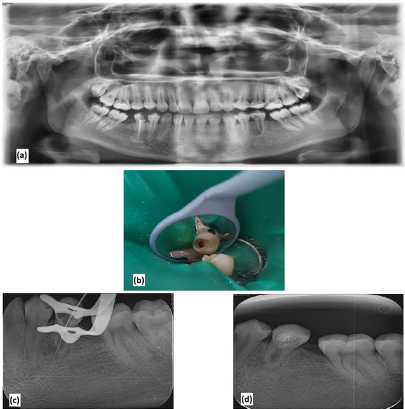
Revitalization of a necrotic mature tooth with external root resorption: Case report and mini review of the literature
Although the conventional apexification procedure—which uses long-term intracanal calcium hydroxide medication to promote the formation of a calcified barrier at the apical foramen—has been widely accepted for more than 40 years, it has significant drawbacks. The weakening effect of prolonged calcium hydroxide application on thin dentinal walls, the resulting high incidence of root fractures, the failure to promote canal wall thickening even when a mineralized barrier is formed using mineral trioxide aggregate, and the unfavorable crown-to-root ratio all contribute to increased susceptibility to fracture. Regenerative endodontic procedures, which apply the tissue-engineering triad of stem cells, biomimetic scaffolds, and growth factors, have therefore been proposed as a biologically based alternative for teeth with pulp necrosis arising from trauma, caries, or developmental anomalies. Although these procedures were developed for immature teeth, protocols employing revascularization and autologous platelet concentrates as scaffolds are increasingly being explored in mature teeth, for which the supporting evidence remains limited. This report describes the regenerative endodontic treatment of an asymptomatic necrotic mature left mandibular second premolar with a chronic apical abscess and external inflammatory root resorption in a 19-year-old woman, using autologous platelet-rich fibrin as a scaffold sealed with mineral trioxide aggregate. Over 12 months of follow-up the tooth remained asymptomatic, the sinus tract resolved, the periapical radiolucency decreased, the resorptive defect appeared arrested, and the tooth responded to thermal testing while remaining unresponsive to electric pulp testing. A focused review of the literature is included to situate this approach among the available alternatives. Within the limitations of a single case with short-term follow-up, this procedure may represent a treatment option for mature necrotic teeth with external inflammatory root resorption; controlled studies with longer follow-up are required.Regenerative endodontics, revitalization, platelet-rich fibrin, root resorption, mineral trioxide aggregateAyşenur Çam, Merve Defişet, Merve Yeniçeri Özata, Anastasiia Bileie250134 -
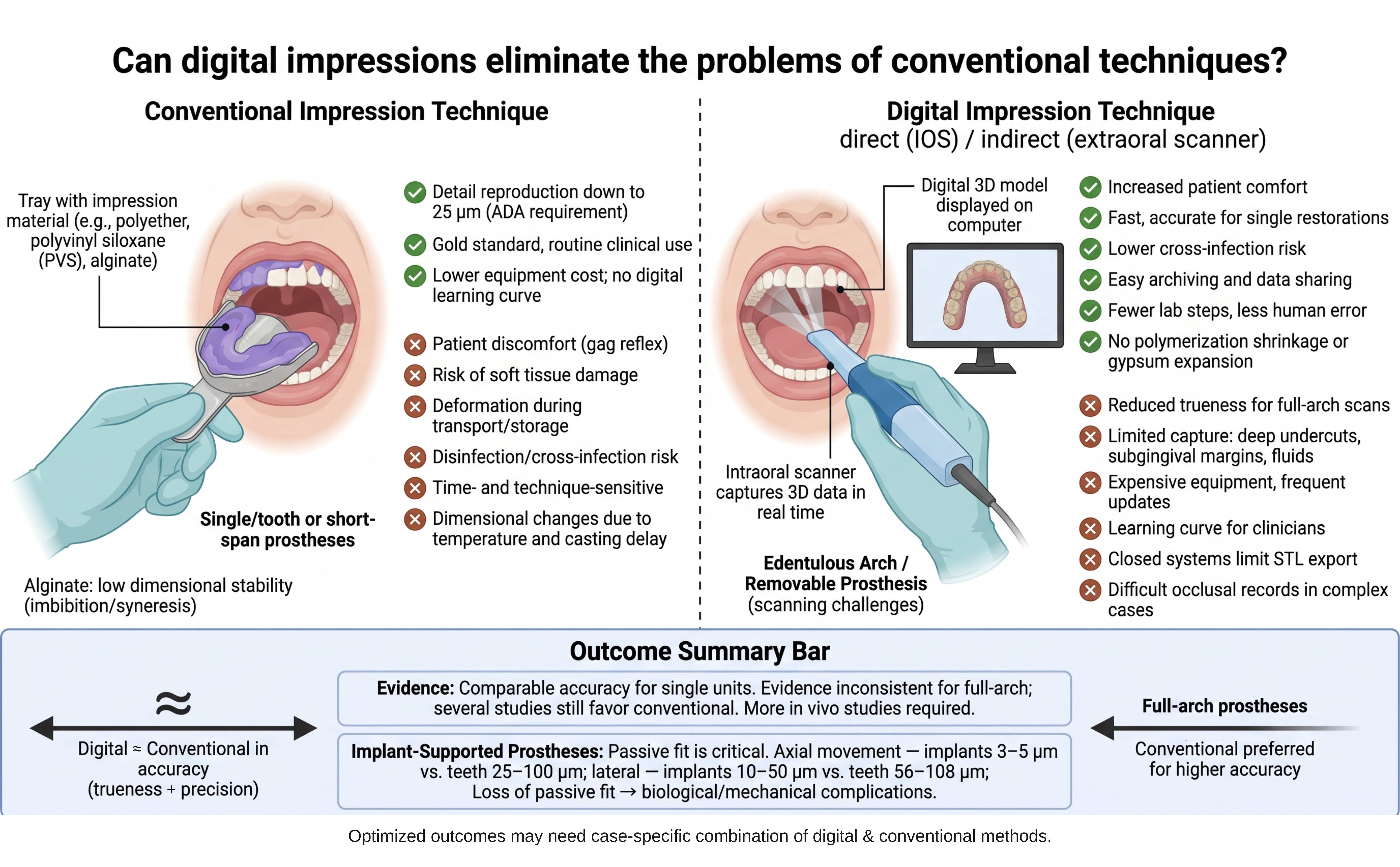
Can digital impression methods eliminate the current problems of conventional impression techniques?
Conventional impression materials remain a cornerstone of routine dental practice, yet they carry well-documented drawbacks, including a pronounced gag reflex, limited patient comfort, potential soft-tissue trauma, dimensional changes with temperature and elapsed time, the need for disinfection, cross-infection risk, and susceptibility to distortion during transport, storage, and laboratory handling. Digital impression systems have been introduced to overcome these limitations, and their use in dentistry—particularly in prosthodontics—continues to expand. This review compares conventional and digital impression techniques and examines whether digital workflows can resolve the shortcomings inherent to conventional methods. Conventional elastomers such as polyether and addition-type silicones offer excellent dimensional stability and detail reproduction and are currently regarded as the gold standard. Digital impressions, obtained with intraoral or extraoral scanners, provide clear advantages in patient comfort, procedure time, reduced cross-infection risk, elimination of polymerization shrinkage and gypsum expansion, and convenient digital archiving and transfer. However, their accuracy is influenced by scanning area, scanner sensitivity, operator experience, moisture, patient movement, and inter-implant distance. The reviewed literature indicates that intraoral scanners achieve clinically acceptable accuracy for single units and single-quadrant restorations, whereas evidence for full-arch and multiple implant-supported prostheses remains inconsistent, with several studies still favoring conventional impressions. Accuracy—comprising trueness and precision—is especially critical for passive fit in implant-supported restorations. Most comparative data derive from in vitro models that do not reproduce intraoral conditions such as saliva, blood, and patient movement, underscoring the need for further in vivo research. In conclusion, although the benefits of digital impressions are evident, their long-term clinical efficacy and safety require additional investigation, and technique selection should weigh accuracy, time efficiency, learning curve, and cost. A case-specific combination of digital and conventional approaches may offer added benefit.Digital impression, intraoral scanner, conventional impression, trueness and precision, prosthodonticsYakup Kantacie260815 -
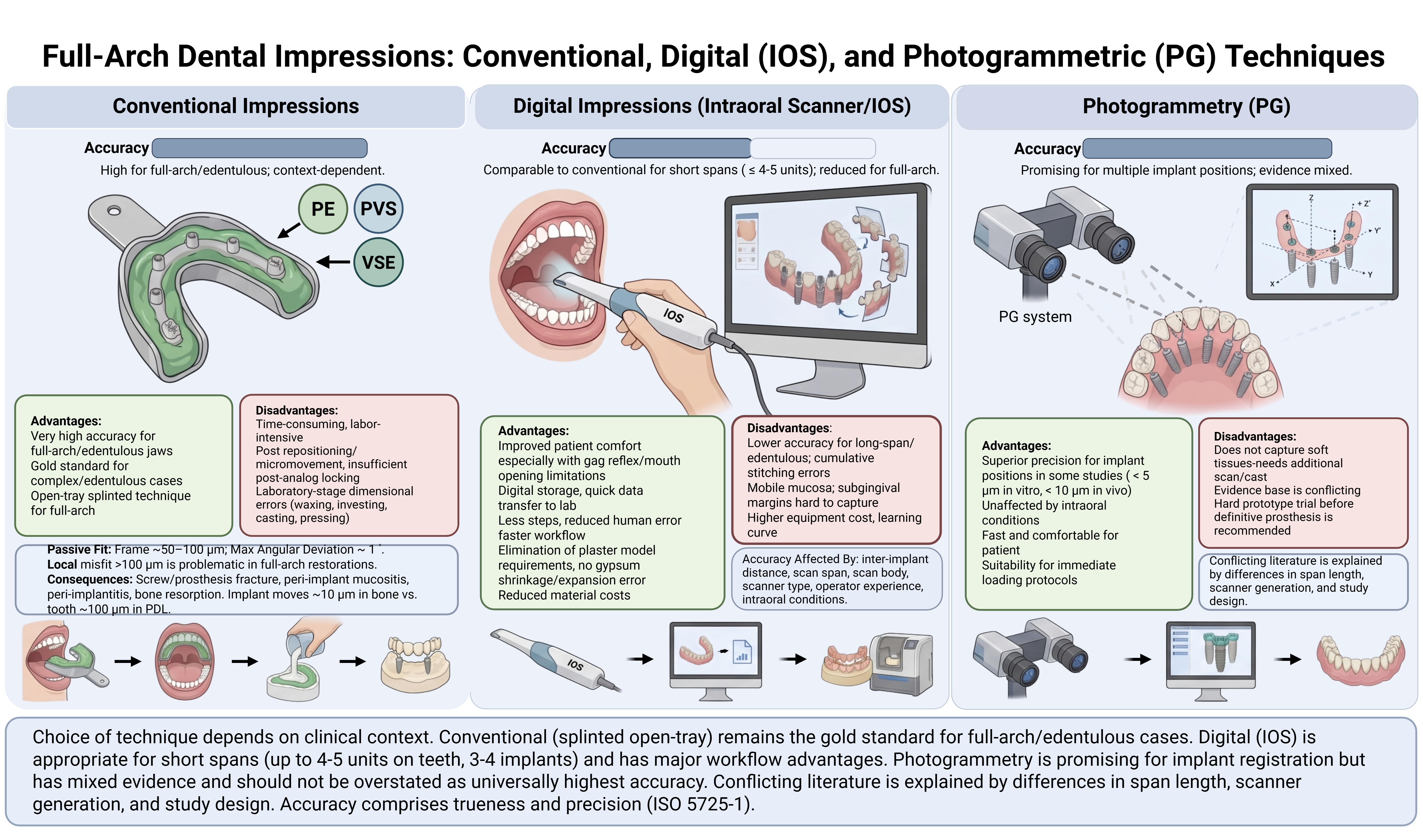
Full-arch dental impressions: Conventional, digital, and photogrammetric techniques
Advances in digital dentistry now permit the fabrication of full-arch prosthetic restorations through a fully digital workflow, extending from impression acquisition to the production of the definitive prosthesis. Intraoral scanning has become an acceptable alternative to conventional impression techniques, providing valuable information for diagnosis, restorative dentistry, orthodontic analysis, and implant-supported prosthodontics. Despite the many advantages offered by digital methods, conventional impression techniques using polyether, polyvinyl siloxane, and vinyl siloxanether materials remain widely used by clinicians because of their excellent dimensional stability and well-documented precision. This review compares and critically discusses conventional and digital impression techniques for acquiring full-arch dental registrations, with particular attention to their accuracy, advantages, limitations, and clinical indications. The accuracy of digital impressions is strongly influenced by inter-implant distance, scan span length, scan body geometry and material, scanner type, operator experience, and intraoral conditions such as mobile mucosa, saliva, and limited anatomical landmarks. For single-unit and short-span restorations of up to four or five units, intraoral scanning achieves accuracy comparable to that of conventional techniques. In long-span and full-arch restorations, however, accuracy tends to decline owing to cumulative image-stitching errors and the scarcity of reference points, a problem that is especially pronounced in completely edentulous arches. Photogrammetry has emerged as a promising alternative for the reliable registration of multiple implant positions, although it inadequately captures peri-implant soft tissues. Overall, technique selection depends primarily on the number of teeth or implants and the distance between them; conventional methods remain preferable for completely edentulous patients, whereas digital workflows are suitable for restorations spanning up to three or four implants. Despite persisting limitations, digital techniques are strong candidates to eventually outperform conventional approaches in dentistry.Full-arch impression, conventional impression, digital impression, intraoral scanner, photogrammetry, implant-supported prosthesis, impression accuracyEbru Sümer Ekine260826 -
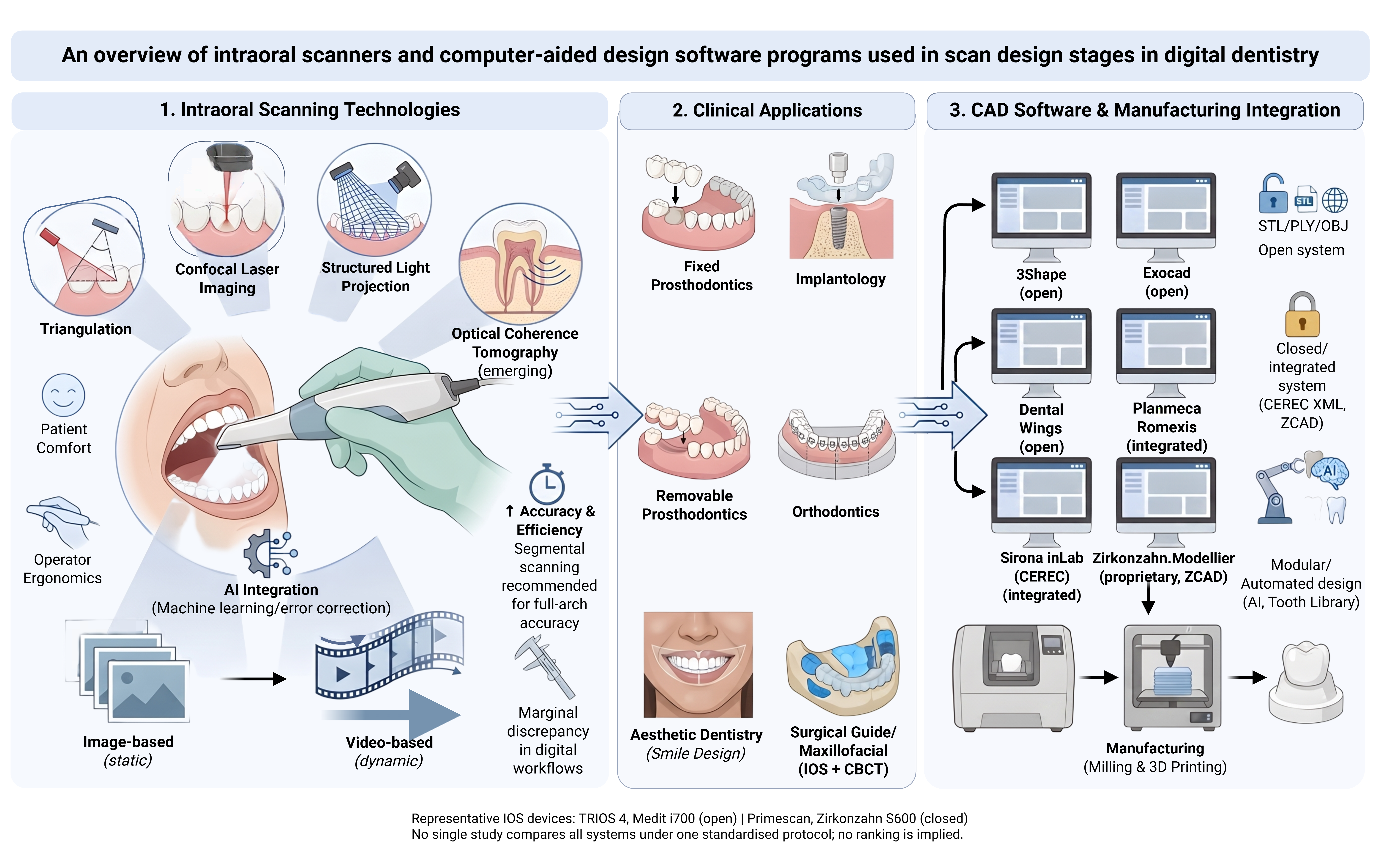
An overview of intraoral scanners and computer-aided design software programs used in scan design stages in digital dentistry
In prosthetic dental treatments, obtaining the negative of the prepared area in the most accurate way directly affects the success of the restoration to be made. Therefore, it is necessary to give importance to the impression material and the impression technique used. Impression materials have undergone changes over the years in order to record the most accurate and sensitive detail. Studies have tried to improve many physical and chemical properties of the impression material. Despite this, it should not be forgotten that there are numerous external factors such as the skill of the clinician, environmental conditions, and storage conditions for the impression to be obtained. Developments in computer-aided design and design (CAD/CAM) technology have significantly increased the use of these systems in dentistry in recent years. In dentistry, many companies in the market aim to minimize the problems that may occur in the laboratory and clinical environment by adding intraoral or extraoral scanners to CAD/CAM systems. The first step of dental CAD-CAM systems is digital scanning with an intraoral or laboratory scanner. This step is followed by the construction phase of restorations with digital design and production devices on the computer. Therefore, the success of these restorations depends largely on the accuracy and precision of the scanning devices. This review will discuss CAD/CAM systems and intraoral scanners and software programs.Digital dentistry, intraoral scanners, CAD/CAM systems, digital impression, scanning accuracy, digital workflowMuhammed Burak Süzen, Hayriye Yasemin Yay Kuşçue260828 -
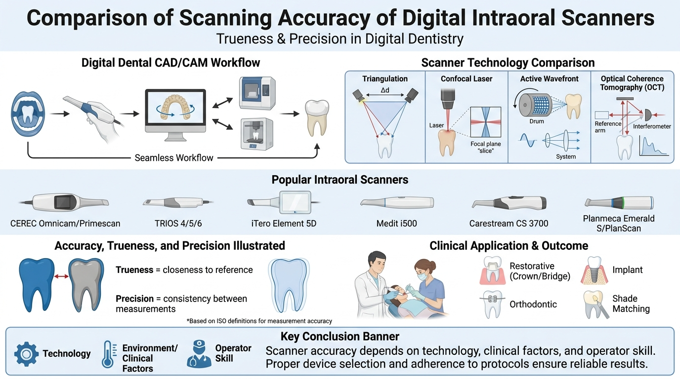
Comparison of scanning accuracy of different digital intraoral scanners
Digital intraoral scanners (IOSs) have gained significant importance in modern dentistry by offering accurate, efficient, and patient-friendly alternatives to conventional impression techniques. This review aims to evaluate and compare the image quality and accuracy provided by various intraoral scanners, focusing on the current literature. Factors influencing image quality, such as scanning technology (confocal microscopy, triangulation, active wavefront sampling), operator experience, and scanning strategy, are also discussed. The evaluation includes commonly used IOS devices, including iTero, Trios, Medit i700, and CEREC Primescan, analyzing their performance in clinical and laboratory conditions. The literature suggests that while each scanner has strengths and limitations, all of them provide clinically acceptable results for prosthodontic and orthodontic applications. Furthermore, advancements in software and hardware are continuously enhancing image resolution, data acquisition speed, and ease of use. However, despite the progress, discrepancies in trueness and precision still exist depending on the scanning environment, scan area size, and material reflectivity. This review concludes that selecting an appropriate IOS should be based on clinical needs, operator preferences, and the specific task at hand. Continuous technological development and standardized evaluation methods are essential to further improve IOS reliability and integration into routine clinical practice.Accuracy, intraoral scanners, digital scanning, three-dimensional imaging, digital dentistryElif Atasever, Server Mutluay Ünale260823 -
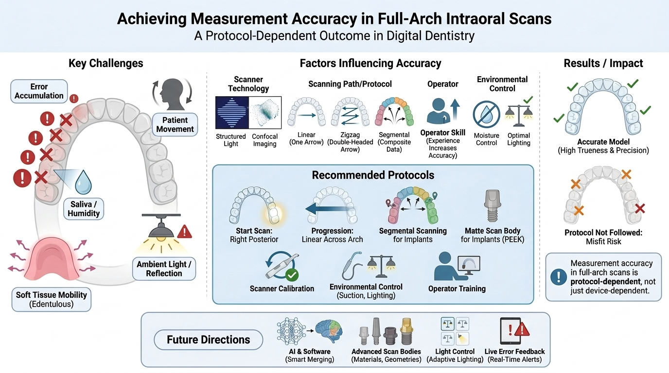
How should I scan for measurement accuracy in full-arch scans?
The integration of intraoral scanners (IOSs) into routine clinical practice has transformed full-arch digital impression-making, offering improved patient comfort and the potential for high measurement accuracy. However, ensuring reliable trueness and precision across large arch spans remains challenging because of the cumulative errors that arise when extended areas are scanned, which can compromise the fit of prosthetic restorations, generate stress within implant-supported frameworks, and introduce clinically relevant errors in orthodontic planning. This review synthesizes the current evidence on the technical, biological, and operator-related factors that influence full-arch scanning accuracy and outlines clinically applicable strategies to minimize error. The principal challenges discussed include local error accumulation, patient movement, saliva and moisture, soft-tissue mobility in edentulous arches, and operator fatigue, all of which can lead to volumetric deformation of the digital model. Key determinants of accuracy are examined, including scanner technology, scanning path and starting-point selection, operator experience, oral humidity, ambient light, and implant scan-body characteristics. Recommended scanning patterns—linear, zigzag, and segmental approaches—are compared and discussed, together with region-specific strategies for the anterior and posterior segments. The evidence indicates that a disciplined protocol markedly improves trueness and precision: linear progression from the posterior toward the anterior region, segmental scanning in implant-supported cases, regular scanner calibration, a constant scanning speed and distance, effective moisture and ambient-light control, the use of matte scan bodies such as PEEK, and continuous operator training. Future research should prioritize intelligent auto-alignment algorithms, standardized implant scan-body geometries and materials, multicenter evaluation of environmental conditions, and artificial-intelligence-based real-time error detection to further enhance the reliability of digital impressions in clinical practice.Full-arch scanning, intraoral scanner, trueness and precision, segmental scanning, digital impressionSavaş Sağmak, Muhammed Burak Süzene260829 -
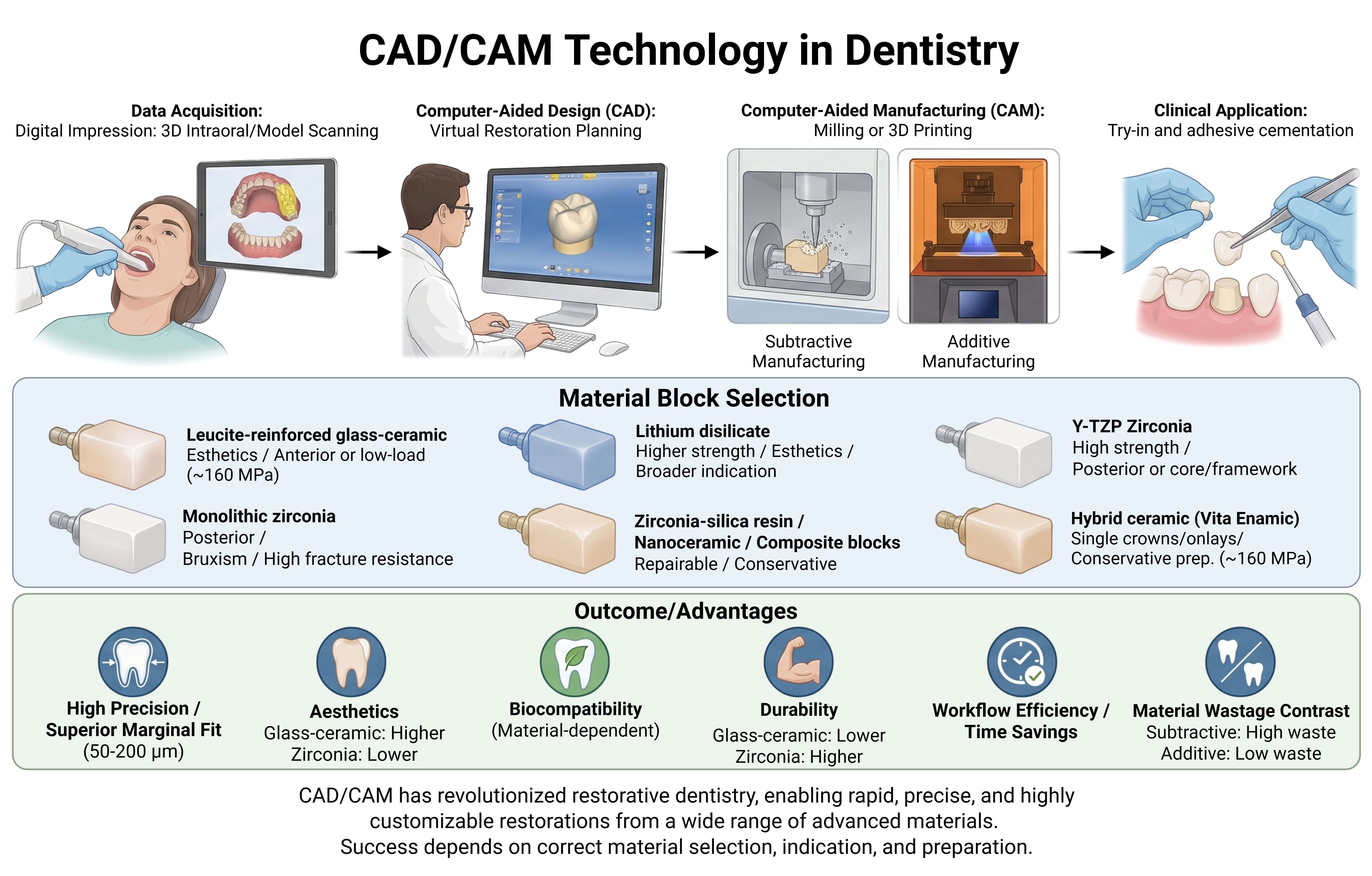
CAD/CAM technology in dentistry
The remarkable advancement of dental materials, treatment techniques, and prosthesis manufacturing processes has elevated computer-aided design and manufacturing (CAD/CAM) systems to a new level of clinical capability. Since its introduction to dentistry in the 1980s—beginning with the chairside CEREC system and the Procera framework concept—CAD/CAM has reshaped contemporary practice by enabling aesthetic, durable, and reliably reproducible restorations while transforming the way laboratories design and fabricate both framework (substructure) and monolithic restorations. Because the production stages are tightly controlled and the material blocks are industrially processed, modern CAD/CAM materials also offer improved structural properties—reduced microporosity, greater homogeneity, and lower firing shrinkage—relative to conventionally processed ones, while reducing laboratory steps, working time, and the overall error rate. This narrative review examines the principal CAD/CAM restorative materials, the digital data-processing workflow, and restoration production methods, together with their influence on marginal fit. The main material classes are described—leucite- and lithium-disilicate-reinforced glass ceramics, yttria-stabilized tetragonal zirconia polycrystals (Y-TZP) and monolithic zirconia, resin-matrix and nanoceramic blocks, composite blocks, and hybrid ceramics such as Vita Enamic—with their compositions, mechanical properties, indications, and limitations, including flexural strengths ranging from approximately 160 MPa for glass ceramics to substantially higher values for zirconia. The data-processing stage, comprising direct intraoral and indirect model scanning followed by computer-aided design of a virtual model, is reviewed, along with the two production routes: subtractive milling from prefabricated blocks and additive fabrication by selective laser sintering and 3D printing, which minimizes material waste. Marginal adaptation is discussed as a key determinant of restorative success, with reported clinically tolerable misfit values of 50 to 200 µm and CAD/CAM milling shown to provide superior marginal conformance to conventional techniques. Overall, CAD/CAM workflows offer measurable gains in material quality, accuracy, and efficiency over conventional methods, although optimal outcomes depend on correct material selection, appropriate indication, and proper tooth preparation.CAD/CAM, dental ceramics, monolithic zirconia, intraoral scanner, marginal adaptation, digital workflowZuhal Görüş, Hayriye Yasemin Yay Kuşçu, Nur Sena Alioğlu Koparale260817 -
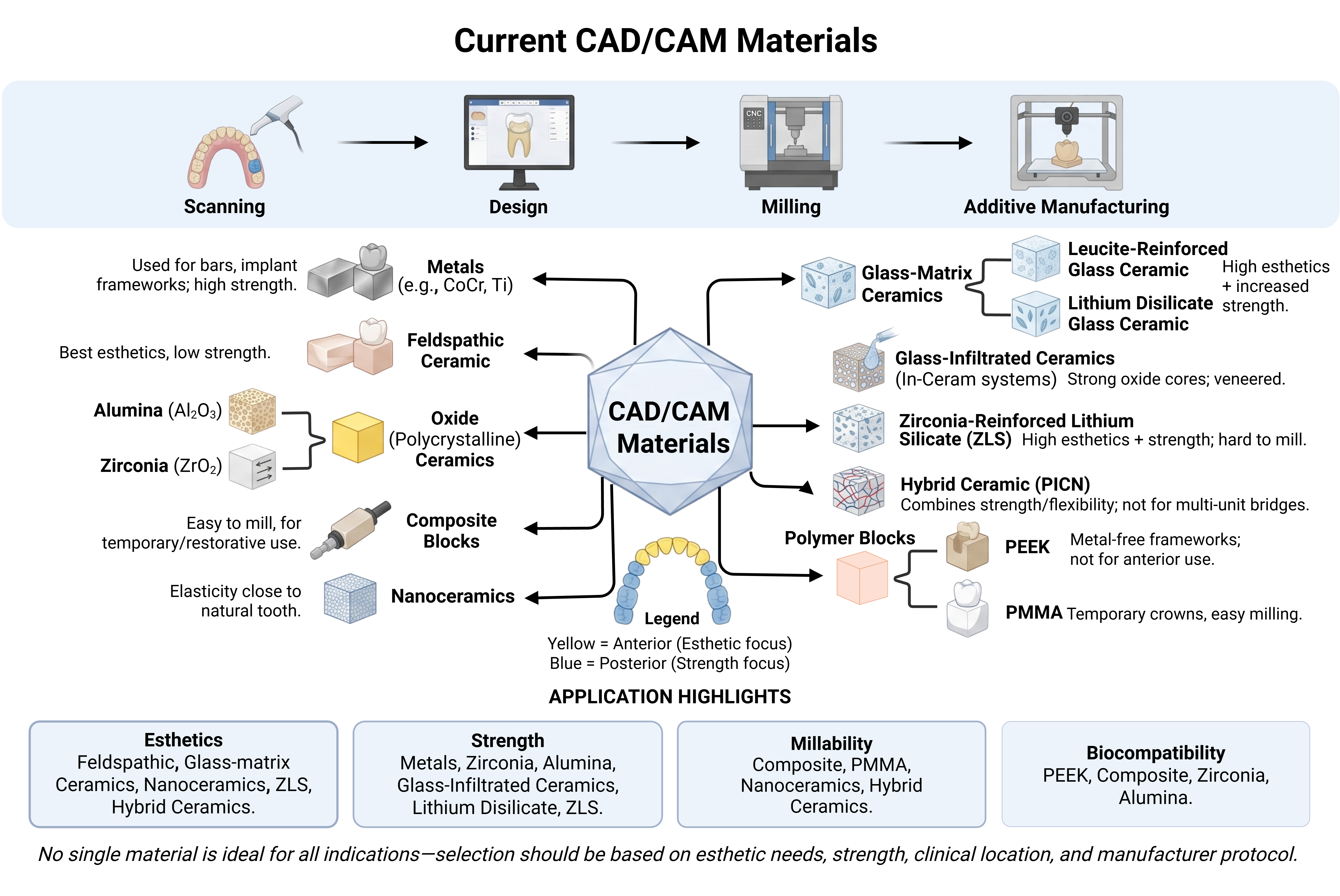
Current CAD/CAM materials
Computer-aided design and computer-aided manufacturing (CAD/CAM) technologies have fundamentally reshaped restorative and prosthetic dentistry, streamlining both chairside and laboratory workflows while improving the speed, accuracy, and predictability of treatment. As the range of millable materials continues to expand, evidence-based, indication-specific selection has become increasingly challenging for clinicians and dental technicians alike. This review classifies and critically evaluates contemporary CAD/CAM materials according to their chemical composition—metal, ceramic, composite, polymer, and hybrid—and compares their performance to support rational treatment planning. Drawing on current literature, it surveys a broad spectrum of materials, including metal alloy blocks; feldspathic, leucite-reinforced, and lithium disilicate glass ceramics; glass-infiltrated and polycrystalline oxide ceramics (alumina and yttria-stabilized zirconia); resin nanoceramics; polymer-infiltrated hybrid ceramics; zirconia-reinforced lithium silicate; composite blocks; and polymer blocks such as PEEK and PMMA. For each group, mechanical strength, optical and esthetic properties, biocompatibility, clinical indications, advantages, and limitations are presented within a comparative framework, and the influence of production strategy—chairside versus centralized fabrication and hard versus soft milling—on restoration fit and longevity is discussed. Reported flexural strength ranged from approximately 150 MPa for feldspathic ceramics to well above 1000 MPa for high-strength zirconia, revealing consistent trade-offs between mechanical performance and translucency, most evident as the cubic phase of zirconia increases. Lithium disilicate and zirconia emerged as leading options balancing esthetics and durability, whereas PEEK and composite materials offered advantages in biocompatibility, elasticity, and provisional applications. Overall, no single material is universally optimal; selection should be guided by the specific clinical indication, esthetic demand, and occlusal load, with ongoing advances in digital dentistry expected to further broaden material choice and clinical performance.CAD/CAM, block materials, metal alloys, ceramics, polymer blocks, digital dentistryPelin Künarcı, Server MUTLUAY ÜNALe260824 -
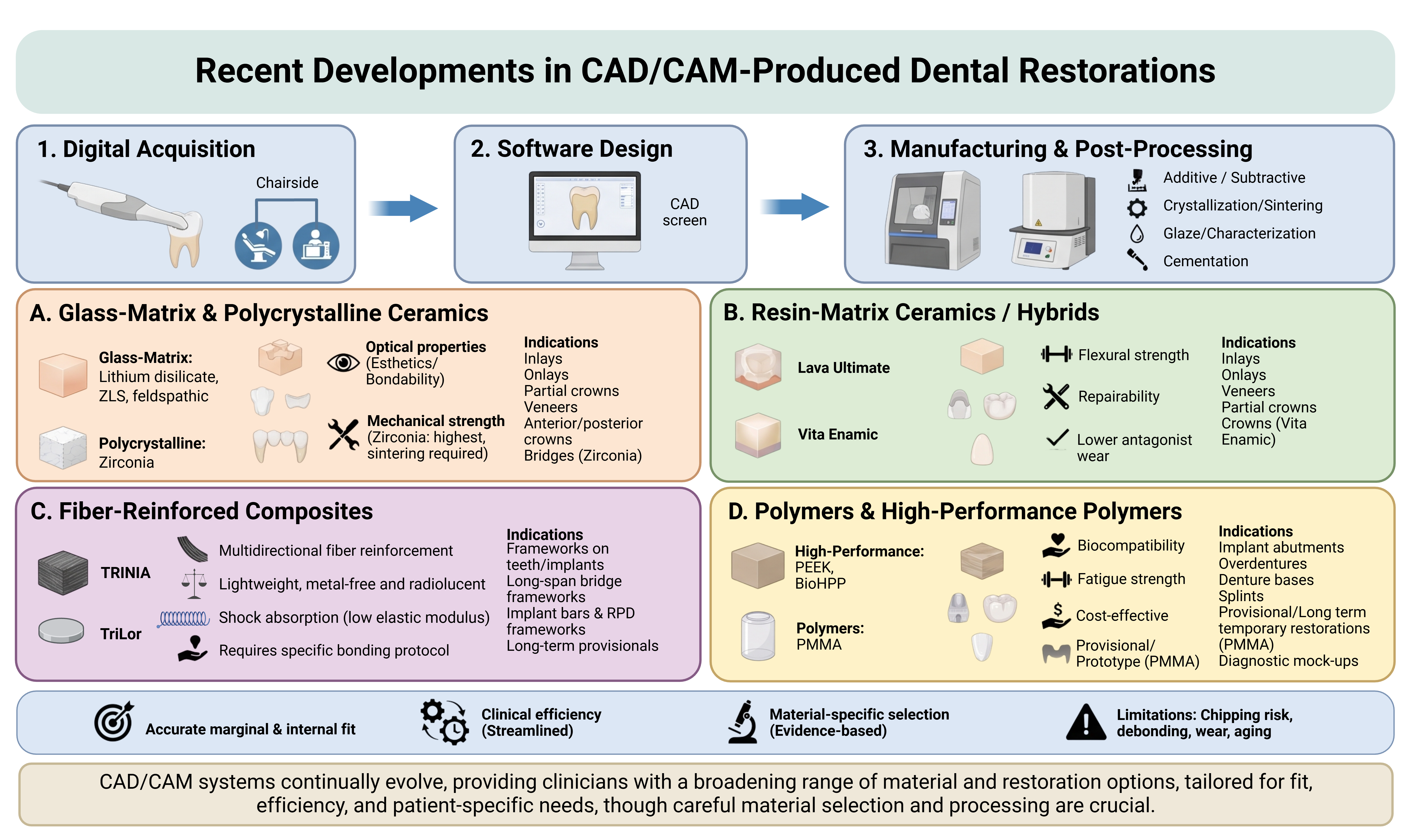
An overview of recent developments in CAD/CAM-produced restorations
The evolution of computer-aided design and manufacturing (CAD/CAM) systems has enabled the development of new materials, treatment methods, and prosthesis manufacturing processes. As innovation in material development continues, the production of new-generation materials with better functions and superior features has become possible. With CAD/CAM technology, chair-side manufacture of esthetic full ceramic restorations that are perfectly compatible with abutments has become possible. In addition, this technology has replaced resin composites applied directly intraorally with resin composite blocks produced with a milling system and applied indirectly, which are mechanically more durable. In addition, digitally produced removable prostheses have also been used widely in recent years. Thus, since the time spent by the dentist for the patient at the clinic and the number of appointments decrease, the delivery time of the prosthesis to the patient is also significantly shortened. Subjective errors due to the technician are also eliminated with these systems, and more accurate manufacture of prostheses is achieved. Because newly developed CAD/CAM materials are manufactured industrially in a standard way and the manufacturing phases are strictly controlled, they can be considered more advantageous than materials used in conventional methods in terms of structural properties. The purpose of this review is to provide physicians with a different perspective on the selection of current materials to be used in the production of CAD/CAM restorations and to assist in material selection in clinical practice.CAD/CAM, glass-matrix ceramics, zirconia, zirconia-reinforced lithium disilicate, fiber-reinforced composites, PEEKMerve Sena Ekinci, Beste Çetine260820






















 Open Access articles are distributed under the terms of the